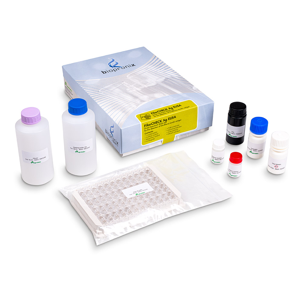
FilarCHECK Ag ELISA
ELISA kit for the detection of Dirofilaria immitis antigen
This kit is based on the monoclonal double antibody ELISA immunoassay technique for the detection of Dirofilaria immitis antigen in canine serum or plasma.
In recent years, our understanding of the pathogenic mechanisms underlying heartworm disease, caused by the parasite Dirofilaria immitis, has undergone a significant evolution. Today, vascular damage is no longer interpreted solely as a mechanical consequence of the presence of adult parasites, friction against the vessel walls or obstruction of blood flow. The latest scientific evidence shows, in fact, that D. immitis is capable of actively interacting with the vascular endothelium, modulating its functions to its own advantage and triggering a complex cascade of immunological and inflammatory events responsible for the development of the clinical disease.
The most recent advances in our understanding of the immunopathogenesis of filariasis are outlined below.
1.Active vascular remodelling: angiogenesis and fibrinolysis
Proliferative endarteritis is the pathognomonic lesion of filariasis, but the mechanisms that trigger it have only recently been elucidated. Through the release of excretory-secretory (ES) antigens, Dirofilaria immitis is able to alter the vascular microenvironment, promoting conditions that enable its survival and contributing to the development of pathological changes.
- Modulation of fibrinolysis: To counteract the formation of clots that might hinder its persistence in the blood vessels, D. immitis secretes proteins (such as actin and fructose-1,6-bisphosphate aldolase) capable of binding to the host's plasminogen, thereby promoting its activation into plasmin. This mechanism represents an advantage for the parasite, as it limits the formation of thrombotic structures around the adult worms. However, chronic stimulation of the fibrinolytic system promotes the degradation of the extracellular matrix and stimulates the proliferation and migration of vascular smooth muscle cells, contributing to the progression of proliferative endarteritis.
- Induction of angiogenesis: The ES antigens of D. immitis can induce the synthesis of pro-angiogenic factors, including vascular endothelial growth factor (VEGF). Persistent activation of the endothelium promotes the formation of abnormal vascular structures resembling pseudo-capillaries and alters the normal permeability of the vessels. Over time, endothelial cells subjected to chronic stimulation may lose their functional organisation, contributing to the pulmonary hypertension associated with cardiopulmonary filariasis.
2.The role of Wolbachia: The 'internal enemy' and the inflammatory response
The discovery of the bacterial endosymbiont Wolbachia pipientis has radically changed our understanding of cardiopulmonary filariasis, highlighting how the pathogenesis of the disease is not determined solely by the nematode, but stems from a complex interaction between three key players: the host, D. immitis and Wolbachia.
- Amplification of endothelial and pulmonary damage: The death of microfilariae or adult parasites, whether naturally or through adulticidal treatment, results in the release of large quantities of Wolbachia and its antigenic components into the bloodstream. These bacterial products, recognized by the host immune system, induce a marked inflammatory response, characterized by the release of pro-inflammatory mediators. This reaction aggravates endothelial and lung tissue damage, promoting local inflammation and increasing the susceptibility to thrombus formation.
- Kidney Damage (Glomerulonephritis): Systemic inflammation induced by Wolbachia and Dirofilaria immitis antigens can cause the formation and deposition of immune complexes in the renal glomeruli, resulting in proteinuria, reduced renal function, and the development of glomerulonephritis, particularly of the membranous type, which can play a crucial role in the prognosis and clinical management of affected patients.
3.The new frontier in staging: serological biomarkers
The evolution of knowledge about the pathogenic mechanisms of heartworm disease has led to the identification of molecular biomarkers as an important aid in disease staging, as they allow for the detection of functional alterations and pathological processes in the early stages, often before they are detectable through traditional imaging techniques such as echocardiography or chest X-rays.
- Endothelin-1 (ET-1): It is one of the most precise markers for assessing the presence and severity of pulmonary hypertension in dogs. Elevated ET-1 levels indicate active vasoconstriction and ongoing remodeling and can persist even months after the parasites have been eliminated.
- NT-proBNP and Troponin I (cTnI): These cardiac biomarkers not only indicate cardiac involvement, but also reflect and provide information on the degree of myocardial stretch (NT-proBNP) and cardiac muscle cell damage/necrosis (Troponin I) caused by right ventricular overload in chronic infections.
- D-dimer and C-reactive protein (CRP): D-dimer has become the parameter of choice for monitoring the risk of pulmonary thromboembolism, especially during adulticidal therapy. CRP, on the other hand, is an important indicator of systemic inflammatory status: its levels correlate with the severity of the disease and the degree of pulmonary hypertension, providing information on the activity of endarteritis and the body's inflammatory response.
Bibliography
- Cardona Machado, C. D., et al. (2023) Involvement of the excretory/secretory and surface-associated antigens of Dirofilaria immitis adult worms in the angiogenic response in an in-vitro endothelial cell model. Veterinary Parasitology, 318, 109939.
- González-Miguel, J., et al. (2015) fibrinolysis and proliferative endarteritis: two related processes in chronic infections? the model of the blood-borne pathogen Dirofilaria immitis. PLOS One, 10(4), e0124445.
- Collado-Cuadrado, M., et al. (2025) Glyceraldehyde 3-phosphate dehydrogenase and galectin from Dirofilaria immitis excretory/secretory antigens activate proangiogenic pathway in in vitro vascular endothelial cell model." Animals (Basel), 15(7), 964.
- Kramer, L., et al. (2008) Wolbachia and its influence on the pathology and immunology of Dirofilaria immitis infection. Veterinary Parasitology, 158(3):191-5.
- Morchón, R., et al. (2012) Anti-Wolbachia surface protein antibodies are present in the urine of dogs with occult Dirofilaria immitis infection with circulating microfilariae but not in dogs with occult infections. Vector Borne Zoonotic Dis. 12(1):17-20.
- Carretón, E., et al. (2017) Cardiopulmonary and inflammatory biomarkers in heartworm disease. Parasites & Vectors, 10(Suppl 2), 534.
- Falcón-Cordón, Y., et al. (2022) Evaluation of acute phase proteins, adiponectin and endothelin-1 to determine vascular damage in dogs with heartworm disease (Dirofilaria immitis), before and after adulticide treatment. Veterinary Parasitology, 309, 109759.
Heartworm disease in dogs generally manifests itself in a very subtle way and the clinical signs are not always easily visible; about four months will pass before the parasites settle in the heart and pulmonary arteries of the animal; then some more time will pass before the parasites reach an amount such as to cause evident symptoms.
Fatigue, poor appetite, difficulty breathing with effort.
Chronic intermittent vomiting also sometimes occurs.
In a more advanced stage we can note:
- weight loss
- increase in respiratory rate
- cough
- erythrocytopenia (reduction in the number of red blood cells)
- increase in abdominal volume
- right heart failure
- epistaxis (nosebleed)
- formation of thromboemboli
- alteration of lung function.