Distemper IC
Rapid test for the detection of Canine Distemper Virus antigen
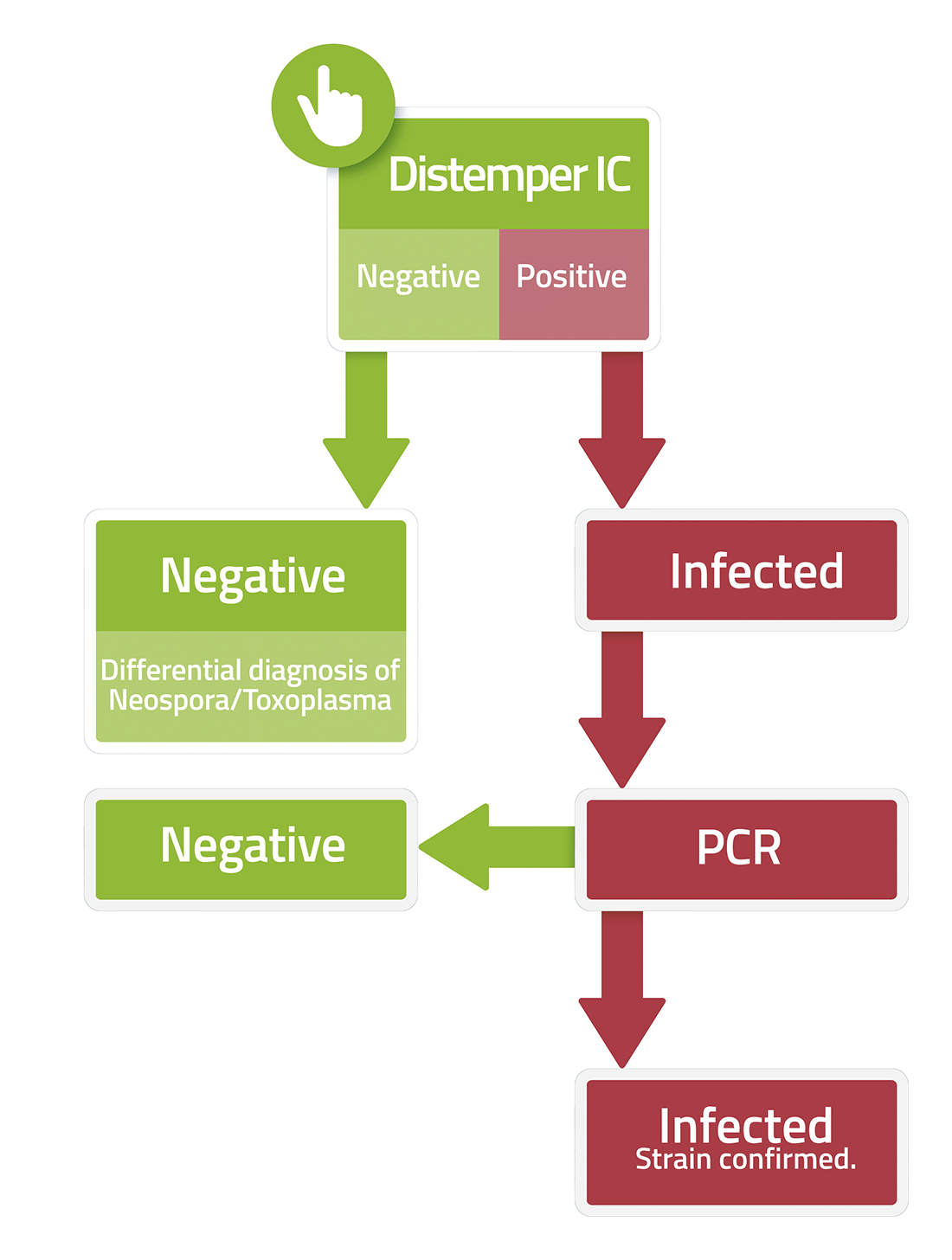
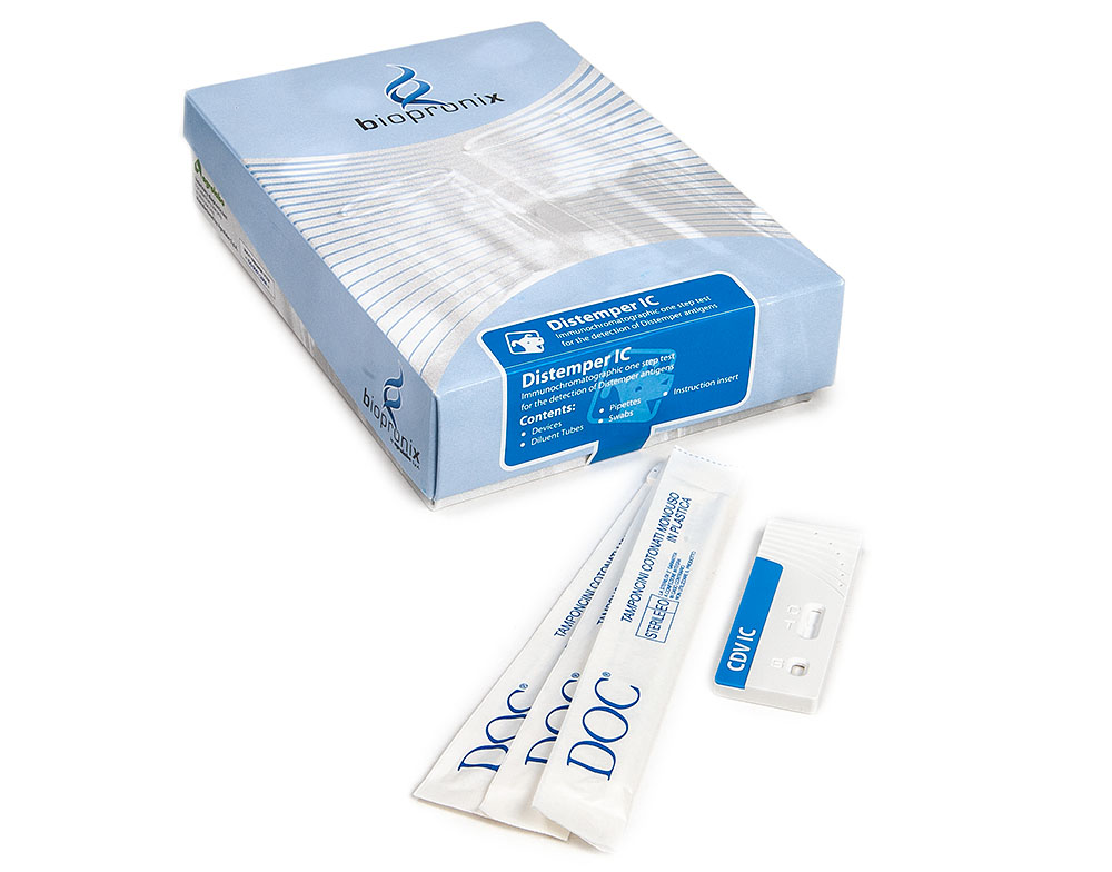
Distemper IC is an immunochromatographic test for the detection of Canine Distemper Virus (CDV) antigen in nasal and conjunctival secretions of dogs
Canine distemper virus (CDV), a member of the Morbillivirus genus of the Paramyxoviridae family, can cause a multisystem disease characterized by respiratory, gastrointestinal, cutaneous, and neurological manifestations. The severity of the infection results not only from the virus's ability to directly damage tissue, but also from its ability to modulate the host's immune system and utilize specific cellular receptors to spread throughout the body.
1.Dual receptor tropism: SLAM/CD150 and Nectin-4
One of the most important aspects of CDV pathogenesis is the virus's ability to utilize two main cellular receptors at different times during infection. This strategy allows the virus to transition from an initial phase characterized by immune compromise to a subsequent phase of dissemination in the epithelium and shedding in the environment.
- First phase: involvement of immune cells through SLAM/CD150. After entering the body through the respiratory tract, CDV primarily exploits the SLAM (Signaling Lymphocyte Activation Molecule, CD150) receptor, expressed on many cells of the immune system, to rapidly reach lymphoid tissues, such as lymph nodes and the spleen, where intense viral replication occurs. The most significant clinical consequence is a marked alteration of the immune response, often associated with lymphopenia, reduced responsiveness to other pathogens, and transient or prolonged immunosuppression.
- Second phase: epithelial spread via Nectin-4. After replicating in immune cells, CDV reaches epithelia using a second receptor, Nectin-4, expressed in the adherens junctions of tissue epithelial cells. Epithelial infection allows the virus to replicate locally and be shed into the environment through respiratory secretions and other biological fluids. Damage to epithelial surfaces contributes to the development of bronchopneumonia, enteritis, skin changes, and hyperkeratosis of the footpads. This phase represents a critical step in the transmission of the virus between susceptible animals.
2.Interferon Evasion: The Role of Proteins V and C
One of the body's first antiviral responses is the production of interferons (IFNs), signaling molecules that activate defense programs in infected and nearby cells. CDV, like other morbilliviruses, possesses sophisticated mechanisms to interfere with this response.
- Interference with the JAK-STAT Pathway: Viral proteins V and C play an important role in modulating the interferon response. In particular, protein V can interfere with STAT1 and STAT2 proteins, key components of the interferon signaling pathway. This results in a reduced ability of the infected cell to activate antiviral genes, communicate the infection status to surrounding cells, and effectively limit viral replication. This mechanism contributes to the rapid spread of the virus in the early stages of infection and to the ability of CDV to reach high viral concentrations in tissues.
3.Syncytia: A Cell-to-Cell Spread Strategy
Like other viruses with fusogenic proteins, CDV uses a unique propagation mechanism through the formation of syncytia.
- Syncytium formation: The viral glycoproteins involved in fusion, particularly the F protein (Fusion protein), allow the membrane of the infected cell to fuse with those of neighboring cells, forming a large multinucleated cell (syncytium) containing viral material from multiple cells.
- Biological advantages: Syncytia promote the direct spread of the virus between adjacent cells, reduce the need for passage into the extracellular space, and partially limit exposure to neutralizing antibodies.
4.Neuroinvasion and Demyelination: Viral Damage and Persistent Inflammation
The neurological manifestations of distemper represent one of the most complex complications of CDV infection. Neurological damage occurs both from viral activity in the central nervous system (CNS) and from the host's inflammatory response.
- Entry into the CNS: CDV can reach the CNS after systemic spread of the infection, including via infected immune cells, which can help transport the virus across biological barriers and facilitate its entry into nervous tissue. Acute phase and damage associated with viral replication. In the early stages of neurological infection, the virus can replicate in CNS cells, including astrocytes, microglia, and other glial cells. Viral replication alters cellular function and causes a local inflammatory process.
- Chronic phase: the role of persistent neuroinflammation. In some cases, even when the amount of detectable virus in the CNS decreases significantly, the demyelination process may continue. This phenomenon is attributed to the persistence of an altered inflammatory response, characterized by prolonged activation of microglia and the production of inflammatory mediators, reactive oxygen species, and enzymes capable of damaging the extracellular matrix and myelin.
Bibliography
- Beineke, A., et al. (2009) Pathogenesis and immunopathology of systemic and nervous canine distemper. Veterinary Immunology and Immunopathology, 127(1-2), 1-18.
- Carvalho, O.V., et al. (2012) Immunopathogenic and neurological mechanisms of canine distemper virus. Adv Virol. 2012:163860.
- Nuhoğlu, H., & Beytut, E., et al. (2026) Immunohistochemical, immunofluorescent, and molecular investigation of Canine Distemper Virus, Nectin 4, and CD150 receptors in naturally infected dogs with Canine Distemper Virus. Animals (Basel). 18;16(14):2233.
- Vandevelde, M., & Zurbriggen, A. (2005) Demyelination in canine distemper virus infection: a review. Acta Neuropathologica, 109(1), 56-68.
- Zhao, J., et al. (2022). Multiple receptors involved in invasion and neuropathogenicity of Canine Distemper Virus: a review. Viruses. 12;14(7):1520.
Distemper is an insidious disease: the symptoms that distinguish it, taken individually, lead back to simple common and not very serious pathologies. The first symptom is generally fever.
The virus causes serious respiratory, gastroenteric, dermatological and neurological problems at a more advanced stage of the disease.
- Respiratory problems: nasal discharge, cough, respiratory distress
- Digestive problems: gastroenteritis with vomiting and diarrhea
- Skin problems: dermatitis, nasal and foot pad hyperkeratosis
- Nervous system problems: convulsions, muscle spasms up to limbs paralysis
- Eye problems: conjunctivitis with ocular drain
.
Following transient leukopenia induced by viral infection, secondary bacterial infection normally manifests as leukocytosis. The transient drop in white blood cells further weakens the immune system and as a result the sick puppy is exposed to secondary infections that complicate the clinical picture.