Ehrlichia IC
Rapid test for the detection of antibodies to Ehrlichia canis
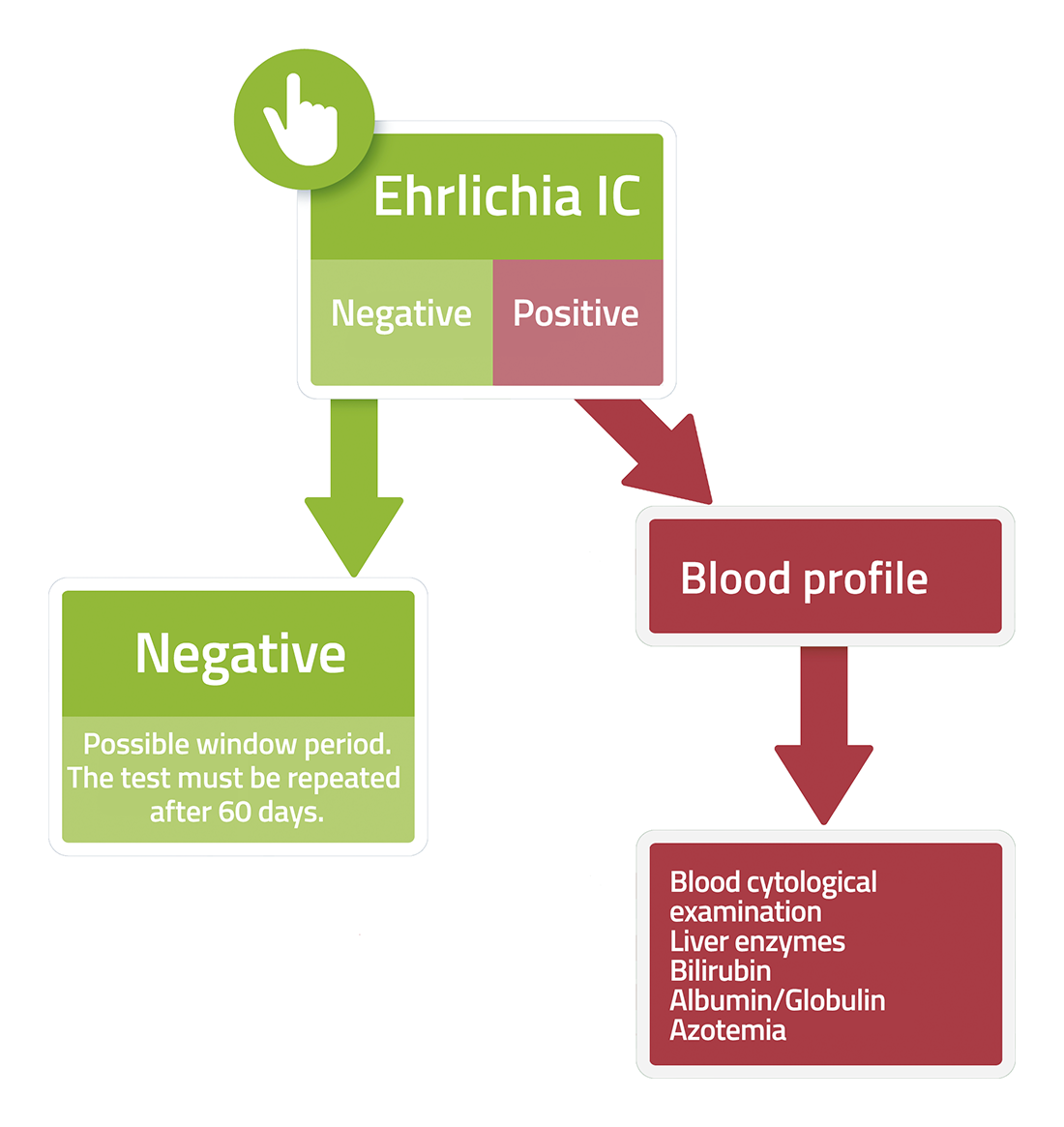
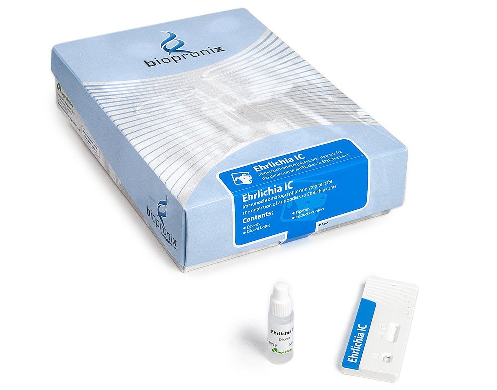
Ehrlichia IC is an immunochromatographic test for the detection of antibodies to Ehrlichia canis in canine whole blood, serum or plasma.
10 TEST
20 TEST
Research has made great strides in understanding Canine Monocytic Ehrlichiosis (CME), caused by Ehrlichia canis. The focus has gradually shifted from simply describing the symptoms (fever, haemorrhages and cytopenia) to understanding the mechanisms by which this obligate intracellular bacterium manages to evade the dog's immune system, ensuring its long-term survival and indirectly contributing to the development of the disease. The most recent and significant advances in our understanding of the immunopathogenesis of ehrlichiosis are outlined below.
1.Phagolysosome Escape and Molecular 'Camouflage'
Ehrlichia canis is a master of deception. Its primary target is monocytes/macrophages, the immune system's scavenger cells that are supposed to destroy it.
- Inhibition of fusion: The latest research has elucidated the molecular mechanisms by which E. canis survives within the host cell. Once phagocytosed, the bacterium inhibits the fusion of the phagosome (a compartment rich in hydrolytic enzymes, reactive oxygen species and other microbicidal molecules designed to eliminate the pathogen) with the lysosome (which contains hydrolytic enzymes, acid hydrolases, capable of degrading bacteria, viruses, proteins, lipids, nucleic acids and other cellular materials). This occurs through the suppression of specific host proteins required for this fusion. The bacterium thus creates a safe 'morula' in which to replicate.
- Silencing the immune system: Ehrlichia canis employs sophisticated immune evasion mechanisms that enable it to limit the activation of the innate immune response. Unlike most Gram-negative bacteria, it does not express detectable levels of lipopolysaccharide (LPS) on its surface and exhibits a marked reduction in peptidoglycan, two of the main pathogen-associated molecular patterns (PAMPs). The absence or low expression of these structures results in reduced activation of pathogen recognition receptors (PRRs), particularly Toll-like receptors (TLRs), thereby attenuating the initiation of the innate inflammatory response. At the same time, E. canis interferes with the antigen-presenting function of infected cells. The bacterium reduces the expression of major histocompatibility complex class II (MHC II) molecules on the surface of monocytes and macrophages, thereby compromising the presentation of antigens to CD4⁺ T lymphocytes. This alteration limits the activation of the adaptive immune response and contributes to the persistence of the infection, as infected cells are recognised and eliminated less efficiently by the immune system.
2.TRP (Tandem Repeat) proteins and the Type 1 secretion system (T1SS)
The latest research into ehrlichiosis focuses on the study of the proteome of Ehrlichia canis and the molecular mechanisms involved in its interaction with the host cell.
- Secret weapons: Several immunogenic proteins have been identified and characterised, including 'Tandem Repeat Proteins' (TRPs), such as TRP19, TRP36 and TRP140. These proteins are substrates of the Type 1 Secretion System (T1SS), a mechanism that Ehrlichia uses to inject its proteins directly into the cytoplasm of the host cell.
- Cellular hacking: Once inside the cell, TRPs and other effector proteins (such as ankyrin repeat proteins – Anks) interact with a vast network of cellular targets, altering gene expression, apoptosis and the monocytic metabolic pathways, transforming the monocyte into a perfect 'incubator'.
- Genetic diversity and virulence: Further studies have highlighted high genetic variability in Ehrlichia canis, particularly in the TRP36 gene, with significant differences between strains isolated from different geographical areas. This evidence suggests that events of mutation and genetic recombination may influence the virulence characteristics of the various strains, contributing to the variability in the clinical presentation of the disease. In particular, certain TRP36 genotypes have been associated with a greater predisposition to haemorrhagic manifestations and more severe clinical forms of the disease.
3.Immunopathology: 'Friendly' damage and the role of the spleen in the immune response
If Ehrlichia canis hides so well, why does the dog fall ill? The answer lies in an exaggerated and misdirected immune response.
- Immune-mediated thrombocytopenia: this is the hallmark of the disease. We now know that the low platelet count is not caused solely by direct destruction by the bacterium. The main mechanisms are increased consumption (due to inflammation-induced damage to the vascular endothelium), splenic sequestration and, above all, immune-mediated destruction. The immune system produces anti-platelet antibodies that cause their premature elimination. Furthermore, the bone marrow undergoes chronic suppression, leading to non-regenerative anaemia and thrombocytopenia in advanced stages.
- The spleen as a 'battlefield': In-depth studies have confirmed that the spleen plays a central role in immunopathogenesis. It is not merely a reservoir where the bacterium persists for years during the subclinical phase. Experimental models have shown that splenectomy (removal of the spleen) significantly reduces the clinical severity of acute disease. This occurs because the main site of antibody synthesis and platelet destruction (phagocytosis by splenic macrophages) is removed.
- The alliance with the vector: A fascinating recent finding concerns the saliva of the Rhipicephalus sanguineus tick. It is not merely a passive vehicle: the saliva contains molecules with anti-inflammatory and immunosuppressive properties that actively inhibit the Th1 response (necessary for eliminating intracellular bacteria) at the bite site, facilitating the initial establishment of E. canis.
These advances shift the focus from organ pathology to the sophisticated molecular mechanisms of host-pathogen interaction.
Bibliography
- Doyle, C. K., et al. (2006) Differentially expressed and secreted major immunoreactive protein orthologs of Ehrlichia canis and E. chaffeensis elicit early antibody responses to epitopes on glycosylated tandem repeats. Infection and Immunity, 74(1), 711-720.
- Francischetti, I. M., et al. (2009) The role of saliva in tick feeding. Frontiers in Bioscience, 14, 2051-2088.
- Harrus, S., et al. (1998) Effect of splenectomy on the course of experimental canine monocytic ehrlichiosis. Veterinary Immunology and Immunopathology, 62(3), 275-282.
- Harrus, S., & Waner, T. (2011) Diagnosis of canine monocytotropic ehrlichiosis (Ehrlichia canis): an overview. The Veterinary Journal, 187(3):292-296.
- Lin, M., & Rikihisa, Y. (2003) Ehrlichia chaffeensis and Anaplasma phagocytophilum lack genes for lipid a biosynthesis and incorporate cholesterol for their survival. Infection and Immunity, 71(9), 5324-5331.
- Luo, T., et al. (2008) Molecular characterization of the Ehrlichia canis TRP36 and E. chaffeensis TRP47 orthologs and identification of an expanded TRP36 ortholog group." Infection and Immunity, 76(4), 1438-1450.
- Rikihisa, Y. (2010) Anaplasma phagocytophilum and Ehrlichia chaffeensis: subversive manipulators of host cells. Nature Reviews Microbiology, 8(5), 328-339.
- Shipov, A., et al. (2008) Macrophage activity in experimental canine monocytic ehrlichiosis. Veterinary Immunology and Immunopathology, 124(1-2), 156-163.
The course of the disease was divided into three phases: acute, subclinical and chronic, based on clinical symptoms and clinical-pathological abnormalities.
The acute phase has a duration of 2 – 4 weeks, during which it is possible to observe symptoms such as:
- fever,
- oculo-nasal discharge,
- anorexia
- depression, lethargy,
- petechiae,
- bruising,
- lymphoadenomegaly in 20% of cases,
- splenomegaly in 25% of cases.
- Most dogs recover from the acute form with appropriate therapy.
Untreated dogs can undergo the subclinical phase.
In this form, body weight normalizes and pyrexia resolves. Dogs appear clinically healthy, however their platelet counts may remain below reference values. Dogs can stay in this phase for years.
The chronic form is characterized by a reduction in hematopoiesis, which can lead to pancytopenia. The prognosis in this phase is poor, death occurs from secondary infections and / or haemorrhage.
Other symptoms that can arise are:
- eye symptoms: change in the color and appearance of the eye, blindness, retinal diseases, anterior uveitis,
- neuromuscular symptoms: meningitis and / or haemorrhagic state, neuropathies, convulsions, atrophic muscles,
- polyarthritis,
- concomitant or secondary infections.