FeLVCHECK Ag ELISA
ELISA kit for the detection of feline leukemia virus antigen
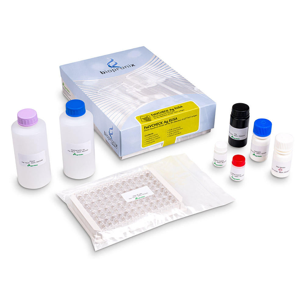
This kit is based on the monoclonal double antibody ELISA enzyme immunoassay technique for the detection of p27 FeLV antigen in felien serum or plasma.
Compared with ViraCHEK/FeLV as reference test, FeLVCHECK Ag agreed at 97.3%, with 97.6% sensitivity (95% confidence interval (CI): 86.0% – 99.9%) and 97.1% specificity (95% CI: 89.1% – 99.5%), while with INgezim FeLV DAS the agreement was 90.2%. Intra- and inter-assay accuracy and precision gave coefficients of variation always lower than 10%.
The new ELISA test FeLVCHECK Ag is a simple and really quick test that permits to identify FeLV Ag in undiluted samples with both visual and spectrophotometric reading.
References:
- A NEW RAPID DIRECT ELISA TEST FOR SEROLOGICAL DIAGNOSIS OF FELINE LEUKEMIA
Irene Ferrero(1), Paolo Poletti(1), Enrica Vittoria Giachino(1), Paola Dall'Ara(2), Joel Filipe(2)
(1) Agrolabo S.p.A, Research And Development, Scarmagno (TO), Italy, (2) Università degli Studi di Milano, Medicina Veterinaria E Scienze Animali, Lodi, Italy – 48th World Small Animal Veterinary Association Congress and 28th FECAVA Eurocongress – Lisbon, 27-29 September 2023 - Feline Leukemia Virus in Cats: A Novel Rapid ELISA Assay for p27 Antigen Detection
Irene Ferrero(1), Paolo Poletti(1), Enrica Giachino(1), Joel Filipe(2), and Paola Dall'Ara(2)
(1)Department of Research and Development, Agrolabo Spa, Scarmagno, Italy – (2) Department of Veterinary Medicine and Animal Sciences, University of Milan, Lodi, Italy
Wiley, Veterinary Medicine International, Volume 2025, Article ID 9914340, 11 pages,
https://doi.org/10.1155/vmi/9914340
Feline Leukaemia Virus (FeLV) is one of the most widely studied pathogenic retroviruses in veterinary medicine. For many years, it was associated primarily with the development of neoplasms and immunosuppression. However, modern advances in molecular virology have revealed a much more complex biological picture. FeLV does not merely infect the host's cells: it integrates its own genetic material into the cat's DNA, establishing a stable relationship with the cellular genome. This characteristic underlies both the persistence of the infection and the mechanisms that may promote the development of haematological and neoplastic diseases.
1. Provirus integration and progressive infection
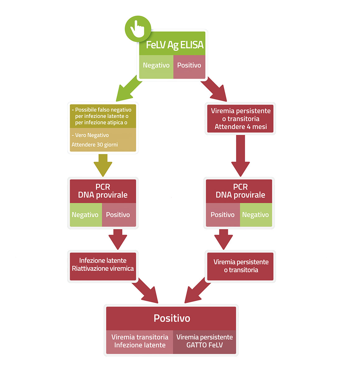
Like all retroviruses, FeLV converts its RNA into DNA through the process of reverse transcription. The viral DNA is subsequently integrated into the genome of the infected cells, forming what is known as a provirus. The course of the infection depends largely on the cat's immune response.
- Progressive infection: When the immune system fails to control viral replication, FeLV spreads to the bone marrow and haematopoietic tissues, leading to persistent viraemia. At this stage, the virus is detectable via the p27 antigen and the risk of developing related diseases is high.
- Regressive infection: Many cats manage to effectively halt viral replication. These cats test negative in standard antigen tests (p27-negative, apparently healthy) and may remain clinically healthy for many years. However, the viral DNA already integrated into the genome is not eliminated. The provirus may persist in a latent form, particularly in bone marrow stem cells, acting as a permanent reservoir of the infection. Under certain conditions, such as severe stress or immunosuppressive treatment, the provirus may reactivate and resume the production of new viral particles, potentially leading to a recurrence of the disease.
2. Insertional mutagenesis: how lymphoma develops
FeLV in its original form does not contain a 'cancer gene' (oncogene). By what mechanism, then, does it cause lymphomas and leukaemias in cats?
- Genome integration: After infecting the cell, FeLV integrates its genetic material into the host's DNA at random. At the ends of the viral genome there are specific sequences, known as LTRs (Long Terminal Repeats), which act as powerful promoters of gene expression.
- Insertional mutagenesis: When integration occurs near genes involved in the control of cell growth, such as the c-myc proto-oncogene, the viral promoters can cause their continuous activation. Consequently, lymphoid cells begin to proliferate uncontrollably, promoting the development of lymphomas and leukaemias. This process is known as insertional mutagenesis and is the main mechanism by which FeLV induces neoplastic transformation.
3. FeLV subtypes and the role of endogenous retroviruses
Natural infection is caused exclusively by FeLV-A, but other viral subtypes may emerge during the course of the disease, arising from genetic changes that occur within the infected organism itself.
- The cat's genome contains sequences of ancient retroviruses, known as endogenous FeLV (enFeLV), which were integrated into feline DNA millions of years ago and are now non-infectious. During viral replication, FeLV-A can recombine with these 'fossil' sequences, giving rise to FeLV-B. FeLV-B has a broader cellular tropism than the original virus and is closely associated with the development of tumours. A different genetic mechanism, however, can generate FeLV-C, a very rare but particularly aggressive subgroup. This virus recognises the FLVCR1 cell receptor, which is essential for the proper metabolism of haem in red blood cell precursors. Interference with this receptor impairs erythroid maturation and can lead to severe aplastic anaemia, characterised by a marked halt in red blood cell production in the bone marrow.
4. The p15E protein and immunosuppression
In addition to its oncogenic effects, FeLV is responsible for a marked immunodeficiency that makes cats more vulnerable to opportunistic infections.
- A central role is played by the viral envelope protein p15E, which not only has a structural function but also possesses potent immunosuppressive activity. The presence of this protein reduces the production of interleukin-2 (IL-2), limits the proliferation of T lymphocytes and alters the normal function of neutrophils. The result is a progressive weakening of the immune response, which promotes the development of secondary infections and contributes to the clinical progression of the disease.
Main Bibliography
- Erbeck, K. et al. (2021) Feline Leukemia Virus (FeLV) endogenous and exogenous recombination events result in multiple FeLV-B subtypes during natural infection. J Virol.95(18):e0035321.
- Fujino, Y., et al. (2008) Molecular pathogenesis of feline leukemia virus-associated malignancies: insertional mutagenesis. Vet Immunol Immunopathol., 123(1-2):138-43.
- Lutz, H. (2009). Feline leukaemia. ABCD guidelines on prevention and management. J Feline Med Surg. 11(7):565-74.
- Overbaugh, J., et al. (2001). Receptors and entry cofactors for retroviruses include single and multiple transmembrane-spanning proteins as well as newly described glycophosphatidylinositol-anchored and secreted proteins. Microbiol Mol Biol Rev. 65(3):371-89
- Quigley, J. G., et al. (2000). Cloning of the cellular receptor for feline leukemia virus subgroup C (FeLV-C), a retrovirus that induces red cell aplasia. 95(3):1093-9
- Tandon, R. (2008) Association between endogenous feline leukemia virus loads and exogenous feline leukemia virus infection in domestic cats. Virus Res. 135(1):136-43.