FIV IC
Rapid Test for the detection of antibodies to Feline Immunodeficiency Virus.
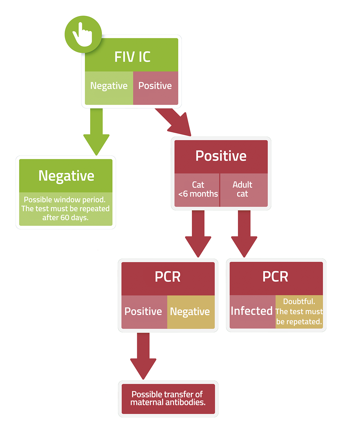
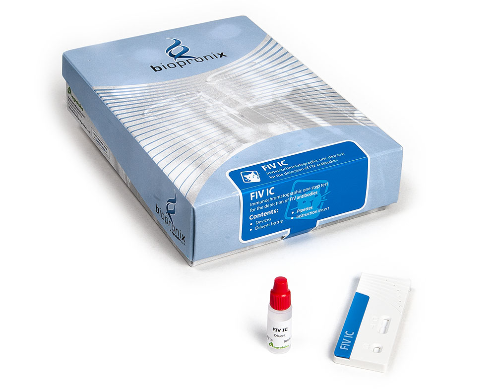
FIV IC is an immunochromatographic test for the detection of anti-FIV antibodies in feline whole blood, serum or plasma.
For many years, the feline immunodeficiency virus (FIV) has been described as the 'feline equivalent of HIV', with the focus primarily on the final stage of the disease, characterised by the progressive impairment of the immune system. However, recent insights into immunopathogenesis have revealed that the virus does not simply act by destroying immune cells: it exploits the very activation of the immune system to replicate and persist within the body. The virus thrives in a state of chronic inflammation, turning the immune response to its own advantage for survival.
1.The role of the CD134 (OX40) and CXCR4 receptors
One of the most significant advances in our understanding of FIV concerns the mechanism by which the virus enters its target cells.
- Unlike HIV, which uses the CD4 receptor, FIV primarily utilises the CD134 (OX40) receptor, which is expressed on T-lymphocytes only when they are activated to respond to an infection. This represents a genuine biological paradox. Whenever a cat faces a common infection, oral inflammation or another immune stimulus, the number of T-lymphocytes expressing CD134 increases. Consequently, the virus has access to a greater number of cells susceptible to infection and can replicate more efficiently. In practice, the more intense the activation of the immune system, the greater the opportunities for FIV to spread
2.T-cell exhaustion and the PD-1/PD-L1 pathway
Why is it that, during the chronic asymptomatic phase, the cat still has T-cells, yet these are unable to eradicate the virus? The answer lies in the phenomenon of 'T-cell exhaustion'.
- Chronic and constant exposure to viral antigens leads T-cells (particularly cytotoxic CD8+ T-cells, which are supposed to kill infected cells) into a state of profound exhaustion and progressive functional inactivity. At a molecular level, these cells express high levels of the PD-1 (Programmed Death-1) receptor. When PD-1 interacts with its ligand PD-L1, an inhibitory signal is transmitted that drastically reduces the immune response. The lymphocytes are not eliminated, but become functionally 'silent': they produce fewer cytokines and lose the ability to destroy infected cells. Understanding the role of the PD-1/PD-L1 pathway represents one of the most significant advances in FIV research, where immune checkpoint inhibitors are used to reactivate the T-cell response.
3.Viral 'reservoirs' and anatomical 'sanctuaries'
One of the main challenges in managing FIV infection is the virus's ability to establish long-term persistence in the body (reservoir).
- Like all retroviruses, FIV converts its genetic material from RNA to DNA and integrates it permanently into the genome of infected cells, forming what is known as a provirus. When this integration occurs in memory T-lymphocytes – cells that are normally quiescent and metabolically inactive – the virus can remain in a state of latency for long periods. At this stage, HIV produces few or no viral proteins, effectively becoming invisible to both the immune system and antiviral treatments. Recent studies have shown that HIV is not found solely in the blood.
- The virus's 'anatomical reservoirs'. The latest research has also shown that FIV is not confined to the bloodstream. The virus is capable of establishing persistent reservoirs within macrophages found in numerous tissues, including the lymph nodes, bone marrow and the gastrointestinal tract. These cells can harbour the virus for years, allowing it to replicate slowly without being destroyed. The presence of these anatomical reservoirs, often referred to as viral reservoirs, contributes to the maintenance of chronic viraemia and is one of the reasons why the complete elimination of the virus remains, to date, an unresolved challenge.
4.NeuroFIV: when inflammation affects the nervous system
In addition to its effects on the immune system, FIV can also affect the central nervous system, causing neurological and behavioural changes known as FIV-associated encephalopathy. This aspect of the disease has made FIV an important experimental model for studying the neurological complications of HIV in humans.
- FIV does not directly cross the blood-brain barrier. To reach the brain, it employs a highly effective strategy: it utilises infected monocytes present in the blood which, once they have migrated into the central nervous system, transform into microglial cells. Once established in the nervous tissue, the virus does not directly infect neurons. The most recent evidence suggests that neurological damage is primarily indirect. Infected microglia and astrocytes release inflammatory mediators, reactive oxygen species (ROS) and neurotoxic substances, such as alterations in glutamate metabolism. This persistent neuro-inflammatory response leads to progressive damage to neurons, contributing to the development of cognitive deficits, behavioural changes and other neurological signs that may occur in FIV-positive cats, particularly in the more advanced stages of the infection.
Main Bibliography
- Brunner, D., et al. (1989) Infection of peritoneal macrophages in vitro and in vivo with feline immunodeficiency virus. J Virol. 63(12):5483-8.
- Fogle, J. E., et al. (2010). PD-1 expression on CD4 and CD8 T cells in feline immunodeficiency virus-infected cats. Veterinary Immunology and Immunopathology, 136(3-4), 305-312.
- Mikkelsen, S.R., et al. (2011) Partial regulatory T cell depletion prior to acute feline immunodeficiency virus infection does not alter disease pathogenesis. PLoS One. 6(2):e17183.
- Podell, M., et al. (2000). "The feline model of neuroAIDS: understanding the progression towards AIDS dementia." Journal of Psychopharmacology, 14(3):205-13.
- Shimojima M., et al. (2004). Use of CD134 as a primary receptor by the feline immunodeficiency virus. Science, 303, 1192–1195.
- Sparger, E.E. (2006) FIV as a model for HIV: an overview. In vivo models of HIV disease and Control. 2006:149–237.
- VandeWoude, S., & Apetrei, C., (2006) Going wild: lessons from naturally occurring T-lymphotropic lentiviruses. Clin Microbiol Rev. 19(4):728-62.
- Willett B. J., et al. (2006). Differential utilization of CD134 as a functional receptor by diverse strains of feline immunodeficiency virus. Journal of Virology, 80(7), 3386–3394.
- Willett, B. J., et al. (2006). Mapping the domains of CD134 as a functional receptor for feline immunodeficiency virus. Journal of Virology, 80(15), 7744–7747.