FLUO PARVOVIRUS
IFA kit for the detection of anti-Canine Parvovirus IgG antibodies
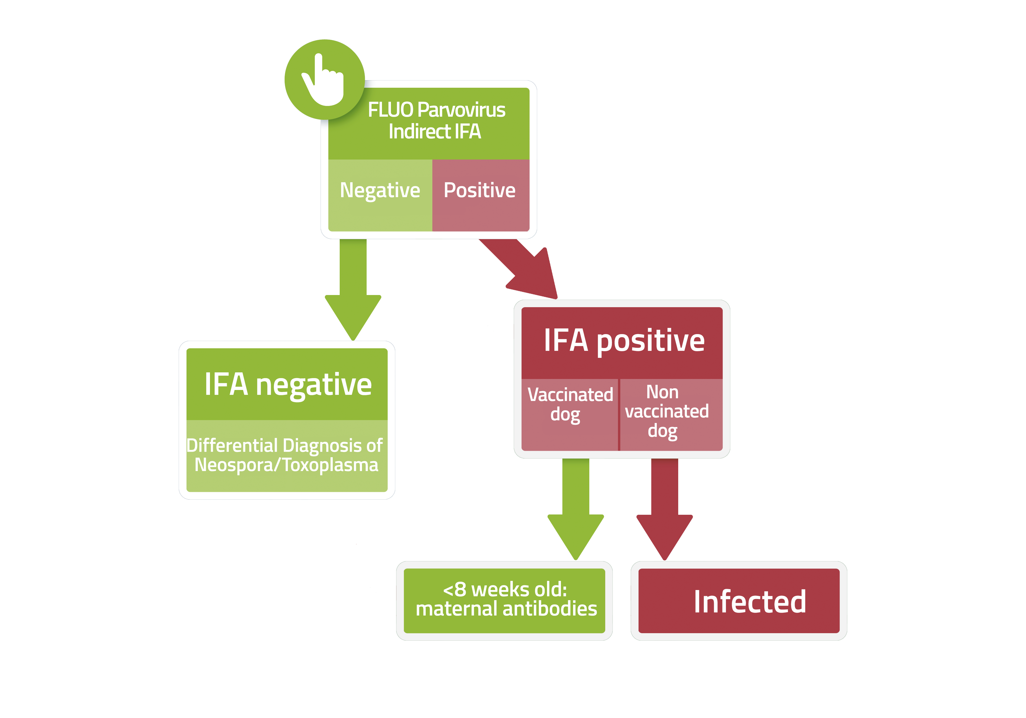
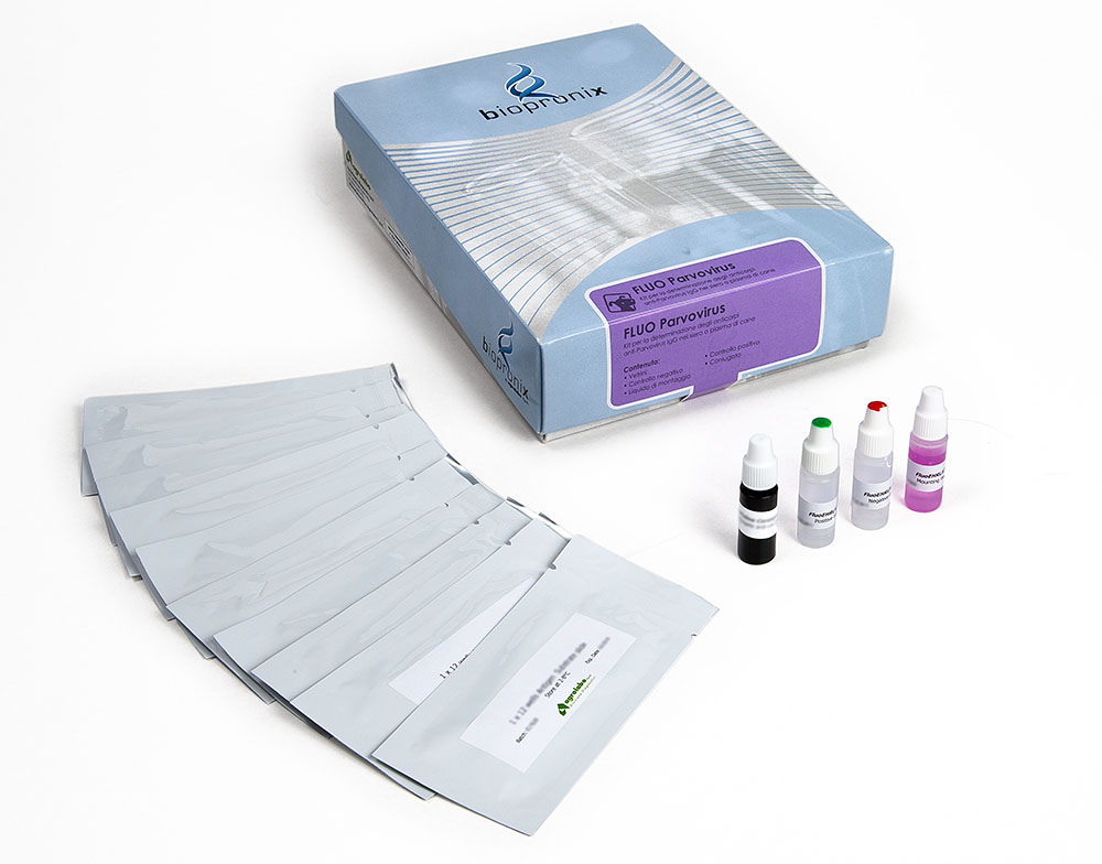
Fluo PARVOVIRUS is a test based on the immunofluorescence technique for the detection of anti-Canine Parvovirus (CPV) IgG antibodies in dog serum or plasma samples.
Canine parvovirus type 2 (CPV-2) is commonly referred to as a simple "enteric virus" responsible for hemorrhagic gastroenteritis. In reality, at the molecular level, CPV-2 is an extraordinarily efficient opportunistic predator and a highly adapted pathogen that not only causes intestinal inflammation but also actively compromises the host's structural and immunological barriers, exploiting the fundamental metabolic and proliferative needs of target cells.
Current knowledge in virology and immunopathology has profoundly altered our understanding of the pathogenic mechanisms responsible for the severity of this infection. The following illustrates the main concepts emerging from the most recent scientific findings.
1.Use of the Transferrin Receptor (TfR) as a Mechanism of Cellular Tropism
CPV-2 is a small, single-stranded DNA virus that does not encode its own DNA polymerase. Consequently, viral replication is strictly dependent on the host cell entering the S phase of the cell cycle, during which the factors necessary for DNA synthesis are available.
- Target cellular receptor: Cells characterized by high proliferative activity (intestinal crypt epithelium, bone marrow hematopoietic progenitors, and neonatal cardiomyocytes) have a marked requirement for iron to sustain cellular replication. For this reason, they express high amounts of Transferrin Receptor 1 (TfR1) on the plasma membrane, which is responsible for the endocytosis of the transferrin-iron complex.
- Virus-receptor interaction: CPV-2 capsid proteins have acquired a high affinity for canine TfR1, which is the main entry receptor of the virus. The evolution from the original strains (CPV-2) to the antigenic variants CPV-2a, CPV-2b, and CPV-2c was driven by point mutations in the VP2 protein, including those located at amino acid residue 426. These substitutions altered the binding properties of TfR1, increasing infectious efficiency in dogs and promoting readaptation to feline transferrin receptors, resulting in a broadening of host tropism.
2.Beyond Diarrhea: Crypts of Lieberkühn, Microbiome, and Translocation
The virus does not cause diarrhea simply by "inflaming" the mucosa. The damage is structural, profound, and architectural.
- Destruction of the Crypts of Lieberkühn: Unlike coronaviruses, which target the tips of the villi (which are more mature and replaceable), CPV-2 infects the germ cells at the base of the intestinal crypts. By killing the "stem" cells, the virus blocks physiological epithelial turnover. The villi collapse, destroying the absorptive surface and the physical barrier of the intestine.
- The Key Role of the Microbiome: The most fascinating studies in recent years have shown that the virus alone is not sufficient to cause the fulminant condition. Experimental models of germ-free dogs (devoid of intestinal flora) exposed to CPV-2 show extremely mild symptoms and survive.
- The Perfect Storm: The true clinical severity is dictated by the preexisting microbiome. When the intestinal barrier collapses, commensal gram-negative bacteria and their endotoxins massively translocate into the bloodstream (endotoxemia). It is secondary sepsis, not the virus itself, that triggers cardiovascular collapse.
3.Enteric Pathogenesis: Crypt Damage, Dysbiosis, and Bacterial Translocation
CPV-2 does not cause diarrhea solely through an inflammatory process of the intestinal mucosa. The damage it causes is predominantly structural, affecting the architectural integrity of the epithelium and the functionality of the intestinal barrier.
- Destruction of the crypts of Lieberkühn: Unlike enteric coronaviruses, which predominantly infect mature enterocytes located at the apex of the intestinal villi, CPV-2 has a marked tropism for the stem and progenitor cells of the crypts of Lieberkühn. The lysis of these cells disrupts physiological epithelial turnover, leading to atrophy and collapse of the villi, resulting in loss of the absorptive surface and impaired mucosal barrier function.
- Role of the intestinal microbiota: Experimental evidence has shown that viral replication alone is not sufficient to cause the most severe clinical manifestations. In germ-free experimental models devoid of intestinal microbiota, CPV-2 infection causes significantly less severe lesions and a significantly more favorable prognosis.
- Pathogenesis of secondary sepsis: The severity of the disease depends largely on the interaction between epithelial damage and intestinal microbiota. Loss of mucosal barrier integrity favors the systemic translocation of commensal bacteria, predominantly Gram-negative, and their lipopolysaccharides (LPS), inducing bacteremia, endotoxemia, and a subsequent systemic inflammatory response. These events are the main determinants of hemodynamic collapse and progression to sepsis.
4.Panleukopenia: Direct lysis and indirect apoptosis
The collapse of white blood cells is the hallmark of the disease, but it does not occur through a single mechanism.
- Lysis in the bone marrow: Again exploiting the TfR, the virus invades the bone marrow and lymphoreticular tissues (thymus, lymph nodes, spleen), replicating within dividing hematopoietic precursor cells and destroying them by direct lysis. The bone marrow literally stops producing new white blood cells.
- Apoptosis of mature lymphocytes: Although mature peripheral T lymphocytes are not actively proliferating and therefore not a direct target of CPV-2 replication, they undergo marked depletion through apoptosis. This phenomenon is not caused by viral lysis, but rather by the activation of apoptotic pathways induced by the systemic inflammatory response and the release of proinflammatory mediators. The resulting lymphopenia amplifies the state of immunosuppression, further compromising the host's ability to contain intestinal bacterial translocation and the progression of secondary sepsis.
5.SIRS and MODS: Pathogenic Mechanisms of Mortality
Death in dogs with CPV-2 infection is not solely attributable to dehydration resulting from diarrhea, but is the result of a complex systemic inflammatory response. Canine parvovirus is currently considered a systemic disease characterized by severe immune dysregulation.
- Activation of innate immunity: Bacterial translocation and endotoxemia resulting from loss of intestinal barrier integrity lead to marked activation of macrophages, monocytes, and endothelial cells through recognition of bacterial lipopolysaccharides.
- Release of proinflammatory mediators: Activation of immune cells induces intense production of proinflammatory cytokines and chemokines, including TNF-α, IL-6, and IL-8, amplifying the systemic inflammatory response.
- Progression to SIRS and MODS: The excessive inflammatory response leads to the onset of Systemic Inflammatory Response Syndrome (SIRS), associated with widespread endothelial dysfunction, alterations in microcirculation and hemostasis, with progression from a prothrombotic state to disseminated intravascular coagulation (DIC). Progression to Multiple Organ Dysfunction Syndrome (MODS) is the leading cause of mortality. Among inflammatory biomarkers, persistently elevated concentrations of C-reactive protein (CRP) are associated with a poor prognosis and an increased risk of adverse clinical outcome.
Bibliography
- Atkinson, B.K. (2022) Circulating markers of endothelial activation in canine parvoviral enteritis. J S Afr Vet Assoc. 93(1):2-7
- Decaro, N., & Buonavoglia, C. (2012). "Canine parvovirus—a review of epidemiological and diagnostic aspects, with emphasis on type 2c." Veterinary Microbiology, 155(1-2), 1-12.
- Goddard, A., & Leisewitz, A. L. (2010) Canine parvovirus." Veterinary Clinics of North America: Small Animal Practice, 40(6), 1041-1053.
- Parker, J. S., et al. (1997). Canine parvovirus host range is determined by the specific conformation of an additional region of the capsid. Journal of Virology, 71(12):9214-22
The course of parvo is variable from subject to subject even within the same litter. There are basically two clinical forms: one gastrointestinal (of variable severity) and one cardiac; however, a sub-clinical form is also recognized.
Gastrointestinal form
After a short incubation period, the following appears:
- vomiting and diarrhea with blood
- anorexia
- depression of the sensory
- dehydration rapidly sets in and, sometimes, even mild hyperthermia
- leukopenia (<3000 cells / mm3) with marked lymphopenia.
.
The course is variable and a slow recovery or a worsening of the general conditions with death of the affected subjects can be observed.
Cardiac form
Clinical evolution is rapid and 2-4 weeks old subjects die with the signs of acute myocarditis.
Subclinical form
It is perhaps the relatively most frequent form, characterized by:
- mild sensory depression
- anorexia
- mild diarrhea
- modest leukopenia / lymphopenia.
.
The development of the sub-clinical form, sometimes asymptomatic, is closely correlated with the degree of immunity of the animal at the time of infection. Obviously, this is a partial immunity that does not prevent the replication and excretion of the virus. This clinical form takes on a different epidemiological importance depending on the kind of life of the animal. In a puppy living in a family environment, the infection will have a limited epidemiological impact; on the contrary, the same situation projected inside a breeding/kennel takes on a completely different implication, as the infected animal eliminates the virus with the feces and can transmit the infection to other subjects.