LeishmaCHECK Ab ELISA
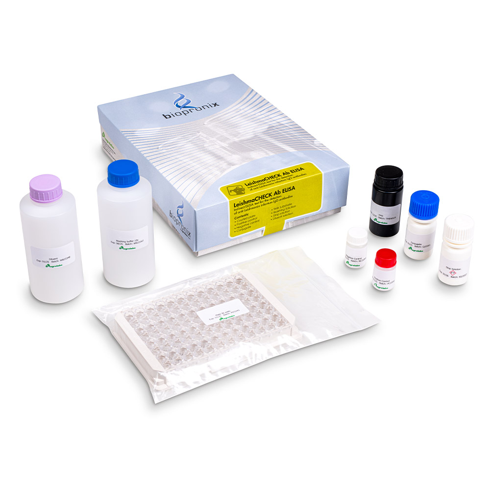
ELISA kit for the detection of anti-Leishmania infantum antibodies
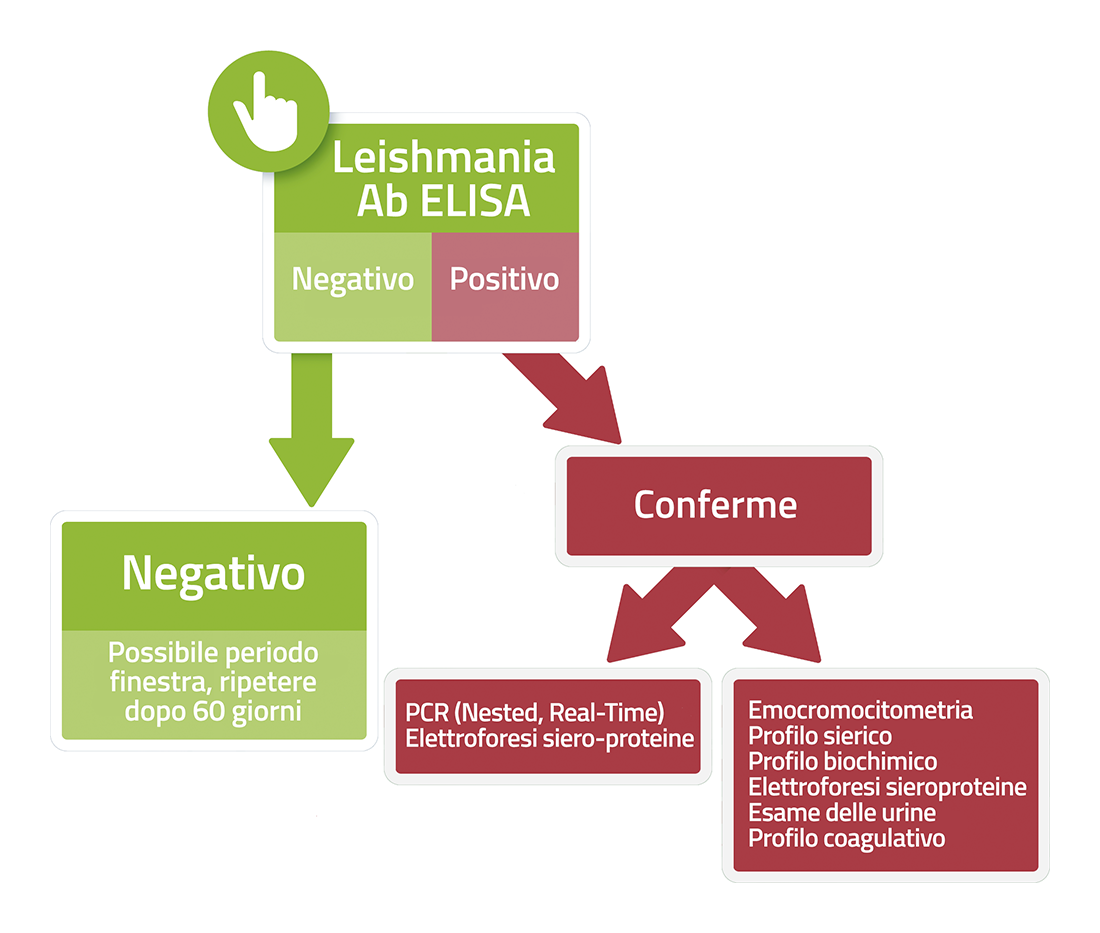
This kit is based on the indirect ELISA immunoassay technique for the detection of anti‑Leishmania infantum antibodies in canine serum or plasma.
The LeishmaCHECK Ab ELISA (Agrolabo) was tested against the reference IFAT test by using 552 sera (333 positives and 219 negatives).
Compared to IFAT as reference test, the new LeishmaCHECK Ab ELISA agrees at 97.1% (Cohen's K: 0.94), sensitivity and specificity are 95.5 and 99.5.
References:
- A new rapid-semi-quantitative indirect ELISA test for the serodiagnosis of Leishmania infantum infection in dogs
Irene Ferrero(1), Paolo Poletti(1), Enrica Giachino(1), Paola Dall'Ara(2), Joel Filipe(2)
(1) Agrolabo SpA, Scarmagno, Italy; (2) Università degli Studi di Milano, Medicina Veterinaria e Scienze Animali, Lodi, Italy
48th World Small Animal Veterinary Association, Congress and 28th FECAVA Eurocongress, Lisbon, 27-29 September 2023 - A newly developed rapid K39-based LeishmaCHECK Ab ELISA for high-throughput serological screening of canine leishmaniasis
Irene Ferrero(a), Paolo Poletti(a), Enrica Giachino(a), Joel Filipe(b) , Alessia Libera Gazzonis (b), Carolina Allievi (b) e Paola Dall'Ara (b)
(a) Dipartimento di Ricerca e Sviluppo, Agrolabo Spa, Scarmagno, Italia
(b) Dipartimento di Medicina Veterinaria e Scienze Animali, Università degli Studi di Milano, Lodi, Italia
https://doi.org/10.1016/j.crpvbd.2026.100418
The modern approach tends to view the dog more as a passive host for the pathogen and focuses on studying the dynamic interaction between Leishmania infantum and the canine immune system, concentrating on the mechanisms that lead to the persistence of the infection or the development of systemic disease.
Below, we describe some of the most recent and significant advances in the immunopathogenesis and pathology of canine leishmaniasis.
1.The central role of circulating immune complexes (CICs)
Whilst the formation of immune complexes has long been recognised, recent studies have focused not only on their presence but also on their active pathogenic role.
In canine leishmaniasis, an uncontrolled infection leads to a massive but ineffective humoral immune response. The combination of high antibody titres (IgG and IgM) and persistent parasitic antigens leads to the formation of circulating immune complexes (CICs).
- Organ damage: CICs precipitate and deposit in the vascular walls of target organs (kidneys, eyes, joints, skin). This deposition is the main cause of the most severe manifestations of canine leishmaniasis, such as glomerulonephritis (often fatal), uveitis, polyarthritis and vasculitis.
- Modulation of the microenvironment: Recent findings suggest that CICs do not merely cause direct mechanical or inflammatory damage. Macrophages activated by these complexes undergo negative modulation: the biosynthesis of IL-12 (crucial for triggering the protective Th1 response and the production of IFN-γ) is inhibited, whilst the secretion of IL-10, an immunosuppressive cytokine, is stimulated.
- Biomarkers of disease severity: New studies have quantified CICs in serum, demonstrating a direct correlation between their levels and molecular size and the progression and clinical severity of the disease (LeishVet staging). This evidence opens up new prospects for the future use of CICs as prognostic biomarkers and as tools for monitoring response to therapy.
2.The role of the immune system: Th1 vs Th2 and regulatory cells
The classification of the Th1 (protective, cell-mediated) and Th2 (humoral, non-protective and potentially pathogenic) immune responses has been refined and expanded following the discovery of the key role played by immunoregulatory cells in the microenvironment of the infection.
- The 'Trojan Horse' effect: Leishmania is a parasite highly specialised in evading the host's defences. Following infection, promastigotes infect macrophages and transform into amastigotes. Instead of being eliminated, the parasites alter the intracellular signalling mechanisms of the macrophages, inhibiting their microbicidal functions—such as the production of nitric oxide—and surviving within the phagolysosomes.
- Regulatory T cells (Tregs) and regulatory B cells: In susceptible dogs, an increase in Tregs and regulatory B cells is observed. These cells actively inhibit the protective Th1 response by producing the pro-inflammatory cytokines IL-10 and TGF-β, thereby promoting the persistence of the parasite. The tissue microenvironment therefore becomes 'tolerant' towards L. infantum, allowing it to survive within the host.
- T-cell exhaustion: Chronic exposure to the parasite's antigens can lead to a state of T-cell 'exhaustion', characterised by reduced proliferative capacity and lower production of inflammatory cytokines (IFN-γ and TNF-α), which are essential for macrophage activation and control of the infection.
3.Non-vector-borne transmission: new epidemiological scenarios
Traditionally linked to the sand fly vector, the pathogenesis and spread of canine leishmaniasis are now also emerging through other evidence, confirmed by recent studies:
- Vertical (transplacental) transmission: Transmission from mothers to puppies during gestation has been extensively documented, including from asymptomatic bitches, in non-endemic areas or in the absence of the vector, confirming that the parasite can be transmitted independently of sand fly bites. This mode of transmission has important implications for the reproductive management of seropositive dogs.
- Sexual and iatrogenic transmission: Although less common, transmission via sexual contact and blood transfusion has been confirmed. For this reason, it is essential to adopt rigorous screening protocols for donor dogs.
4.New frontiers in diagnosis: beyond conventional serology
Advances in our understanding of the immunopathogenesis of canine leishmaniasis have led to the development of increasingly accurate diagnostic tools, capable of improving the early identification of infection and supporting more effective clinical management.
- Recombinant antigens: Modern molecular biology techniques have enabled the identification and production of highly specific recombinant antigens for L. infantum. The use of these antigens in rapid immunochromatographic tests is significantly improving field diagnosis, reducing cross-reactions and enhancing the early identification of infected animals.
- Molecular diagnostics (PCR): PCR techniques, in particular real-time PCR, performed on target tissues (bone marrow, lymph nodes, skin and conjunctiva), are essential for detecting even low parasite loads, identifying asymptomatic dogs and confirming infection in cases where serology is inconclusive or negative, such as in the early stages of the disease or in dogs with a severely compromised immune response.
Advances in scientific knowledge confirm that canine leishmaniasis is a complex condition: not merely an infectious disease, but an immune-mediated disorder in which the interaction between the parasite and the host's immune system plays a decisive role.
Bibliography
- Boggiatto, P. M., et al. (2011) Transplacental transmission of Leishmania infantum as a means for continued disease incidence in North America. PLoS Negl. Trop. Dis. 5(4):e1019.
- Cortese, L., et al. (2013) Regulatory T cells, cytotoxic T lymphocytes and a T(h)1 cytokine profile in dogs naturally infected by Leishmania infantum. Vet. Sci. 95(3):942−9.
- Costa, F. A., et al. (2003) Histopathologic patterns of nephropathy in naturally acquired canine visceral leishmaniasis. Veterinary Pathology, 40(6): 677–684.
- Esch, K.J., Petersen, C.A. (2013) Transmission and epidemiology of zoonotic protozoal diseases of companion animals. Microbiol. Rev. 26(1):58−85.
- Latrofa, M. et al. (2016) Vertical transmission of Anaplasma platys and Leishmania infantum in dogs during the first half of gestation. Parasites & Vectors, 9, 269.
- Maia, C., & Campino, L. (2008) Methods for diagnosis of canine leishmaniasis and immune response to infection." Veterinary Parasitology, 158(4), 274–287.
- Paltrinieri, S., et al. (2016). Laboratory tests for diagnosing and monitoring canine leishmaniasis. Vet. Clin. 45(4):552−578.
- Petersen, C. A., & Barr, S. C. (2009) Canine leishmaniasis in North America: emerging or newly recognized?. Veterinary Clinics of North America: Small Animal Practice, 39(6), 1065–1074.
- Solano-Gallego, L., et al. (2011). LeishVet guidelines for the practical management of canine leishmaniosis. Parasites & Vectors, 4(1), 86.
- Toepp, A.J., & Petersen, C.A. (2020) The balancing act: Immunology of leishmaniosis. Vet. Sci. 130:19−25.
Symptoms of cutaneous leishmaniasis:
- Dry exfoliative dermatitis and progressive alopecia around the eyes, on the legs and on the back. Dermatitis can degenerate and cause lesions and ulcers – even without itching – usually affecting the eyes, ears, nose and mucous membranes; in severe cases, blood may also leak from these.
- Ulcers can cause swelling of the lymph nodes (in 90% of cases),
- Accelerated growth and abnormal thickening of the nails (onychogryphosis) (in 20% of cases).
- Joint pain and lameness including back pain: the dog often stands motionless, keeping his head down to seek relief (in 37.5% of cases).
Symptoms of visceral leishmaniasis:
- renal failure, polydipsia, polyuria (in 40% of cases)
- vomiting and diarrhea, which result in loss of appetite and rapid and evident weight loss (in 64% of cases)
- neurological damage or even uremic coma
- nosebleeds (epistaxis) due to ulcers in the oral mucosa, in which parasites are present (in 15% of cases)
- ocular lesions, due to a uveitis and iridocyclitis and conjunctivitis (respectively in 1.3% and 32.5% of cases).