Lyme IC
Rapid test for the detection of antibodies to Borrelia burgdorferi
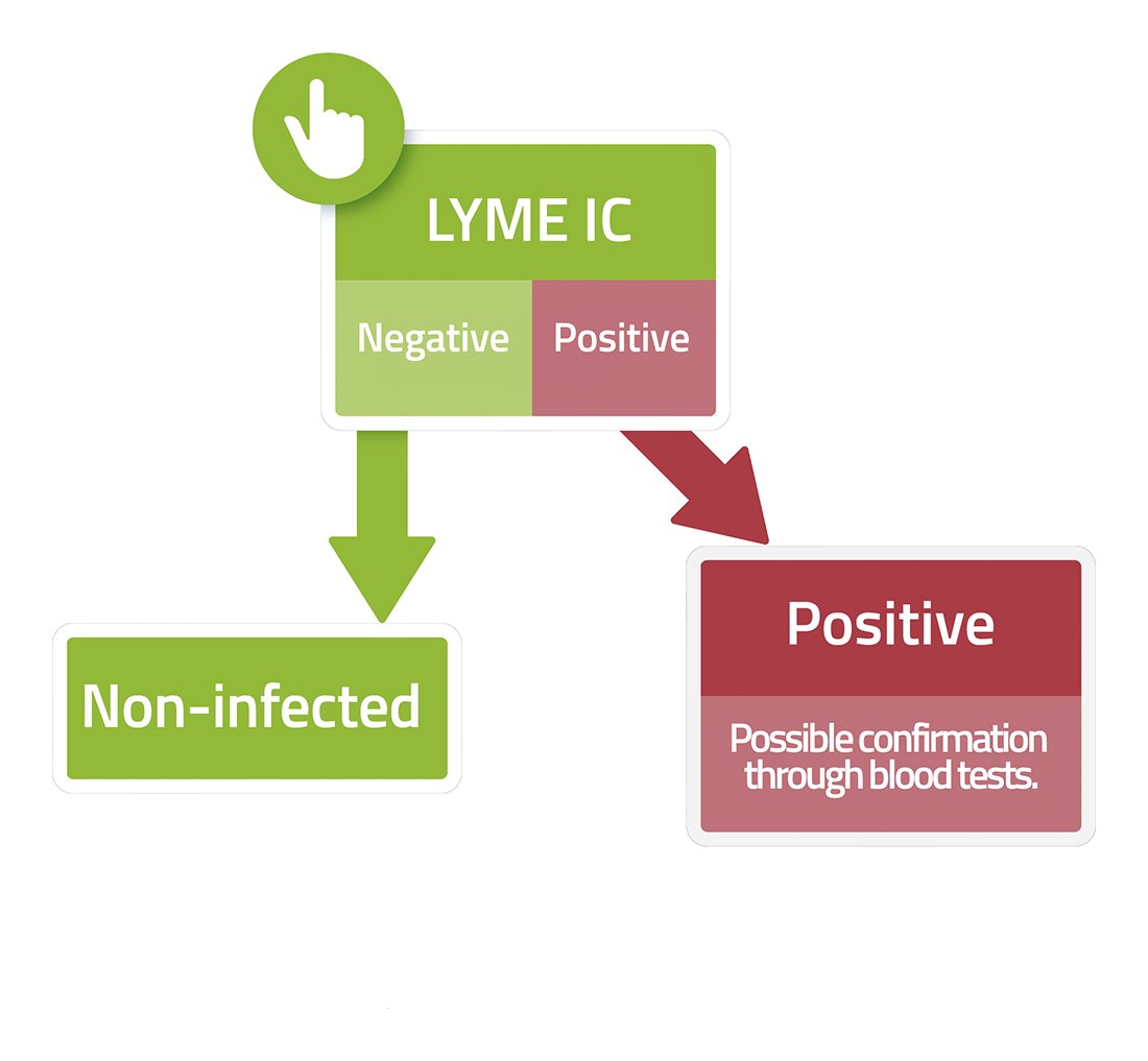
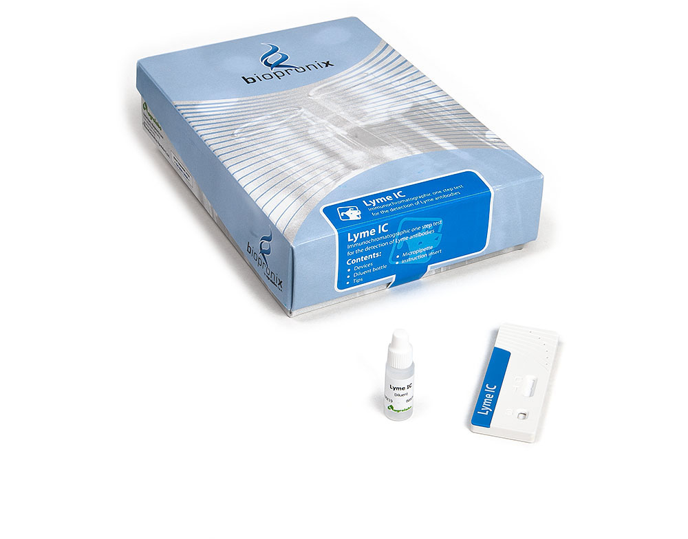
Lyme IC is an immunochromatographic test for the detection of antibodies to Borrelia burgdorferi in canine whole blood, serum or plasma.
Lyme disease, caused by spirochetes belonging to the Borrelia burgdorferi sensu lato complex, represents one of the most studied examples of evolutionary adaptation between a microorganism, its arthropod vector, and its mammalian host. In addition to the clinical manifestations observed in dogs, such as intermittent lameness associated with arthritis, the pathogenesis of Lyme disease is characterized by sophisticated molecular mechanisms that allow the bacterium to rapidly adapt to the diverse biological environments encountered during its life cycle.
The most recent scientific findings have highlighted the fundamental role of surface protein regulation and antigenic variation in allowing Borrelia to colonize mammals and persist despite the host's immune response.
1.Molecular transition: the OspA–OspC system
The transfer of Borrelia from ticks to dogs requires a complex modification of the bacterium's gene expression.
- OspA: In ticks that have not yet engaged in a blood meal, Borrelia predominantly express OspA (Outer Surface Protein A), a surface protein involved in the bacterium's adhesion to the gut cells of the vector Ixodes spp. This phase allows the microorganism to maintain its niche within the tick until the next blood meal.
- Blood meal and OspC: During the tick's meal, increased temperature and environmental changes associated with the presence of the mammal's blood activate a regulatory cascade in the bacterium. A reduction in OspA expression and an increase in OspC (Outer Surface Protein C) production are observed.
- OspC and the initial phase of infection: OspC is essential for the migration of Borrelia from the tick's gut to the salivary glands and contributes to the bacterium's ability to establish infection early after transmission. This protein interacts with various host components and promotes the initial survival of the microorganism before an effective adaptive immune response develops.
2.VlsE and Antigenic Variation: The Mechanism of Immune Persistence
Once in the dog's body, Borrelia faces strong selective pressure from host-produced antibodies. The primary immune evasion mechanism is the VlsE (Vmp-like sequence Expressed) system, responsible for the continuous variation of the surface proteins expressed by the bacterium.
- Genetic recombination and bacterial surface modification: The vlsE gene undergoes frequent genetic recombination events, progressively modifying the variable regions of the VlsE protein exposed on the bacterium's surface. This ability allows Borrelia to rapidly generate new antigenic variants, reducing the effectiveness of antibodies previously produced by the host.
- Persistence of infection: The dog's immune response can partially control the infection, but continuous antigenic variation allows a population of bacteria to escape antibody neutralization and maintain tissue persistence. This phenomenon is one of the main reasons why the natural immune response does not always result in complete elimination of the bacterium.
- C6 peptide as a diagnostic marker: The C6 peptide derives from the unchanged region of the VlsE protein and is widely used in modern serological tests for the diagnosis of exposure to Borrelia burgdorferi. Its diagnostic importance stems from its high conservation among different Borrelia strains and its ability to stimulate a specific antibody response during infection.
3.Complement Evasion: The Role of CRASP Proteins in Borrelia Survival
The complement system is an essential component of innate immunity and one of the first defenses against microorganisms present in the blood and tissues. Through a complex enzymatic cascade, complement can cause pathogen opsonization, the recruitment of immune cells, and the formation of the Membrane Attack Complex (MAC), responsible for perforating the cell membrane and killing the microorganism.
To survive in the host, Borrelia burgdorferi has developed sophisticated mechanisms for regulating complement activation, which are essential for the persistence of the infection.
- CRASP Proteins and Factor H Recruitment: Among the main immune evasion tools of Borrelia are CRASPs (Complement Regulator-Acquiring Surface Proteins), proteins expressed on the bacterial surface that can interact with complement regulators present in the host's plasma. In particular, some CRASPs can bind Factor H and FHL-1 (Factor H-like protein 1), molecules normally involved in controlling the activation of the alternative complement pathway on the body's cells.
- Reduction of complement activity: The recruitment of regulatory factors to the bacterial surface allows Borrelia to reduce uncontrolled complement activation and limit the deposition of terminal components of the cascade, including the MAC. This mechanism does not represent true "camouflage" as a host cell, but rather an effective molecular strategy that allows the bacterium to increase its resistance to the innate immune response.
4.Lyme disease-associated nephropathy: an immune-mediated complication
Although the most well-known clinical manifestation of canine borreliosis is Lyme polyarthritis, one of the complications is Lyme disease-associated nephropathy. This condition is rare, but can cause a serious clinical picture characterized by proteinuria, progressive loss of renal function, and, in the most severe cases, renal failure.
- Immune-mediated damage: The renal damage associated with borreliosis is generally immune-mediated, attributable to a type III hypersensitivity reaction, in which the antibody response leads to the formation of antigen-antibody complexes.
- Immune complexes and glomerular damage: The persistence of Borrelia antigenic components, combined with the host immune response, can promote the formation of circulating immune complexes (CICs). When these complexes deposit in the renal glomeruli, they can activate complement and attract inflammatory cells such as neutrophils and macrophages, causing damage to the glomerular filtration barrier, resulting in membranoproliferative glomerulonephritis, marked proteinuria (often fatal), and acute renal failure.
Bibliography
- Dambach, D. M., et al. (1997) Morphologic, immunohistochemical, and ultrastructural characterization of a distinctive renal lesion in dogs putatively associated with Borrelia burgdorferi infection: 49 cases (1987-1992). Vet Pathol. 34(2):85-96.
- Kraiczy, P., et al. (2003) Immune evasion of Borrelia burgdorferi: mapping of a complement-inhibitor factor H-binding site of BbCRASP-3, a novel member of the Erp protein family. Eur J Immunol. 33(3):697-707.
- Liang, F. T., et al. (1999). An immunodominant conserved region within the variable domain of VlsE, the variable surface antigen of Borrelia burgdorferi. The Journal of Immunology, 163(10), 5566-5573.
- Littman, M. P. (2013) Lyme nephritis. Journal of Veterinary Emergency and Critical Care, 23(2), 163-173.
- Schwan, T. G., et al. (1995) Induction of an outer surface protein on Borrelia burgdorferi during tick feeding. Proceedings of the National Academy of Sciences, 92(7), 2909-2913.
- Tilly, K., et al. (2008) Biology of infection with Borrelia burgdorferi. Infectious Disease Clinics of North America, 22(2), 217-234.
- Zhang, J. R., et al. (1997) Antigenic variation in Lyme disease borreliae by promiscuous recombination of VMP-like sequence cassettes. Cell, 89(2), 275-285.
- fever 39.5-40.5 °C,
- intermittent lameness affecting the different limbs,
- joint swelling,
- lymphoadenomegaly,
- anorexia and general discomfort,
- polyarthritis usually in the limb closest to the tick attack site,
- nephropathies,
- meningitis,
- initial transient skin rash that develops where the tick has stung the animal.