MalasseziaKit
Rapid microbiological test for the identification of Malassezia.
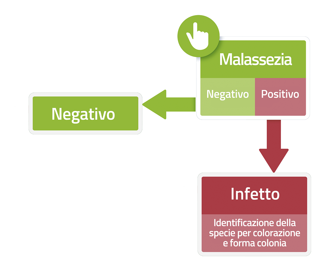
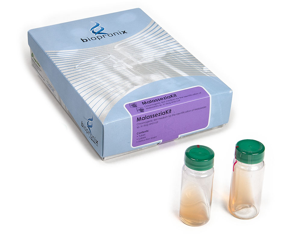
MALASSEZIAKIT is a rapid microbiological test for the identification of Malassezia of different species in samples of dog and cat hair and/or earwax.
Skin and ear cytology is the primary diagnostic tool for evaluating Malassezia infections in dogs and cats. Simple and rapid techniques, such as tape stripping, skin impressions, or ear swabs, allow for the detection of yeasts and prompt treatment.
However, in patients with chronic, recurrent, or poorly responsive dermatitis or otitis, cytology alone may not provide all the information needed for optimal management. In these settings, microbiological testing by culture and, when indicated, species identification and assessment of antifungal susceptibility represent an important diagnostic complement.
1.Species-level identification
Cytology allows for the documentation of the presence of yeasts compatible with the Malassezia genus, but does not allow for a definitive species identification. For many years, Malassezia pachydermatis, a non-lipid-dependent species, was thought to be the only one of real clinical interest in companion animals.
- Lipid-dependent species: Modern microbiological culture methods using specific media (such as the MalasseziaKit test medium) have demonstrated that, especially in cats and, more rarely, in dogs, other Malassezia species can also be isolated, including some lipid-dependent species, such as M. sympodialis, M. furfur, M. globosa, and M. nana.
- Clinical Impact: Species-level identification can be achieved by culture on selective media and, when necessary, confirmed with advanced microbiological identification methods. Species-level characterization can be useful in selected cases, especially when the clinical course is atypical or the therapeutic response is unsatisfactory.
2.From simple isolation to strain characterization
The presence of Malassezia on the skin does not necessarily equate to disease. These yeasts are part of the normal skin microbiota, and their pathological proliferation depends on the interaction between the microorganism, the skin barrier, and numerous predisposing factors, such as allergies, endocrine disorders, or alterations in the skin environment.
- Virulence factors: Modern microbiological techniques allow for detailed study of some biological characteristics of isolated strains, including the production of extracellular enzymes (lipases, phospholipases, proteinases). These virulence factors contribute to the alteration of the epidermal barrier, local inflammation, and the persistence of skin lesions.
- Biofilm: a possible cause of chronic forms. Recent microbiological studies have demonstrated that M. pachydermatis is able to organize itself into a biofilm, an extracellular matrix produced by microorganisms that adheres to the skin surface or ear canal and in which the yeasts remain embedded. This structure may limit the penetration of antifungals, increase tolerance to treatments, promote the persistence of the infection, and contribute to clinical relapses.
Bibliography
- Álvarez-Pérez, S., et al. (2024) Prevalence and genetic diversity of azole-resistant Malassezia pachydermatis isolates from canine otitis and dermatitis: A 2-year study. Med Mycol. 62(5):myae053.
- Bond, R., et al. (2020) Biology, diagnosis, and treatment of Malassezia dermatitis in dogs and cats. Clinical Consensus Guidelines of the World Association for Veterinary Dermatology. Vet Dermatol. 31(1):28-74.
- Cafarchia, C., et al. (2012) In vitro antifungal susceptibility of Malassezia pachydermatis from dogs with and without skin lesions. Veterinary Microbiology, 155(2-4), 395-398.
- Figueredo, L.A., et al. (2012) Biofilm formation of Malassezia pachydermatis from dogs. Vet Microbiol. 160(1-2):126-31.
- Guillot, J., et al. (1998) Usefulness of modified Dixon's medium for quantitative culture of Malassezia species from canine skin. J Vet Diagnostics Invest. 10(4):384-6.
In veterinary dermatological diagnostics, cytology remains the primary tool for identifying yeasts using rapid stains, such as Diff-Quik® or methylene blue. However, for further microbiological investigation, culture is the traditional method for confirming the growth of the microorganism and obtaining more information about its identity.
The introduction of chromogenic culture media represented a major diagnostic advance. Unlike common cytological stains, these media exploit specific biochemical reactions to differentiate microorganisms during growth, offering rapid and easily interpretable presumptive identification.
1.Color as an indicator of metabolic activity
In traditional culture media, such as Sabouraud Dextrose Agar (SDA), Malassezia colonies generally present a similar appearance, with a creamy-white color and poorly distinguishable morphology.
- Chromogenic media: They contain chromogenic substrates that are metabolized by specific enzymes produced by microorganisms. The hydrolysis of these substrates releases colored compounds that accumulate in the colony, allowing visual distinction between different metabolic profiles.
- Color for interpreting results: Colony color is a biochemical indicator, useful for guiding the identification of the most frequently isolated species in veterinary medicine. It is important to emphasize that color is a presumptive identification criterion and must be interpreted in the context of morphological and clinical characteristics and, when necessary, confirmed using advanced identification methods.
2.Greater ability to recognize mixed cultures
One of the main advantages of chromogenic media is the ability to highlight the simultaneous presence of microorganisms with different metabolic characteristics.
- Conventional cultures: Colonies belonging to different species may present a very similar appearance, or predominant growth may mask less represented microorganisms.
- Chromogenic medium: Any biochemical/enzymatic differences can result in the appearance of colonies with different colorations, facilitating the recognition of a possible mixed culture. This type of use represents an important support for the microbiologist, as the presence of different species can influence clinical interpretation and guide further diagnostic investigations.
3.Reduced decision-making times
Traditionally, identifying Malassezia species required various methods, such as mass spectrometry (MALDI-TOF MS) or molecular biology techniques, and specific biochemical tests.
- Rapid diagnosis: The use of a chromogenic medium allows for a first indication of the possible identity of the microorganism after just 48–72 hours of incubation, reducing the time required for microbiological assessment.
Bibliography
- Cabañes, F. J. (2014). Malassezia yeasts: how many species infect humans and animals?. PLoS Pathogens, 10(2):e1003892.
- Cafarchia, C., et al. (2011) Advances in the identification of Malassezia. Mol Cell Probes. 2011 Feb;25(1):1-7.
- Kaneko, T., et al. (2007) Revised culture-based system for identification of Malassezia species. J Clin Microbiol. 45(11):3737-42.
This Dermatitis is caused by a yeast belonging to the genus Malassezia. All Malassezia species are lipo-dependent, with the exception of Malassezia pachydermatis, which is generally lipophilic. Malassezia pachydermatis is the most commonly found species in mucous membranes and on the skin of cats and especially dogs.
These yeasts are a part of the normal cutaneous microflora and their overgrowth can become pathogenic under certain conditions that lead to a change in the cutaneous microclimate and favour their replication, which causes external otitis and severe itching dermatitis. Malassezia dermatitis is typically itchy and erythematous; secondary lesions such as alopecia, excoriations, seborrheic plaques, lichenification, are common and in these cases, it is not possible to distinguish a Malassezia dermatitis from a staphylococcal pyoderma without a specific examination.
Dermatitis may be localized (ear, face, interdigital spaces, anal area) or generalized.
Malassezia otitis causes itching ears and head shaking. Ear wax is abundant and has a brownish consistency, with a strong rancid smell.
The diagnosis is complicated because Malassezia infection often occurs in association with other diseases according which the differential diagnosis is carried out: atopy, flea allergy, adverse food reactions, superficial pyoderma, external parasites, neoplasms, endocrinopathies and other metabolic disorders that can promote its overgrowth. Other predisposing factors consist of breed (dogs: WHWT, Basset Hound, Maltese, Cocker, Poodle; cats: Sphynx, Devon Rex, Persian), humid climate and warm seasons, presence of skin folds and alterations of the stratum corneum.