FLUO CANINE HERPESVIRUS
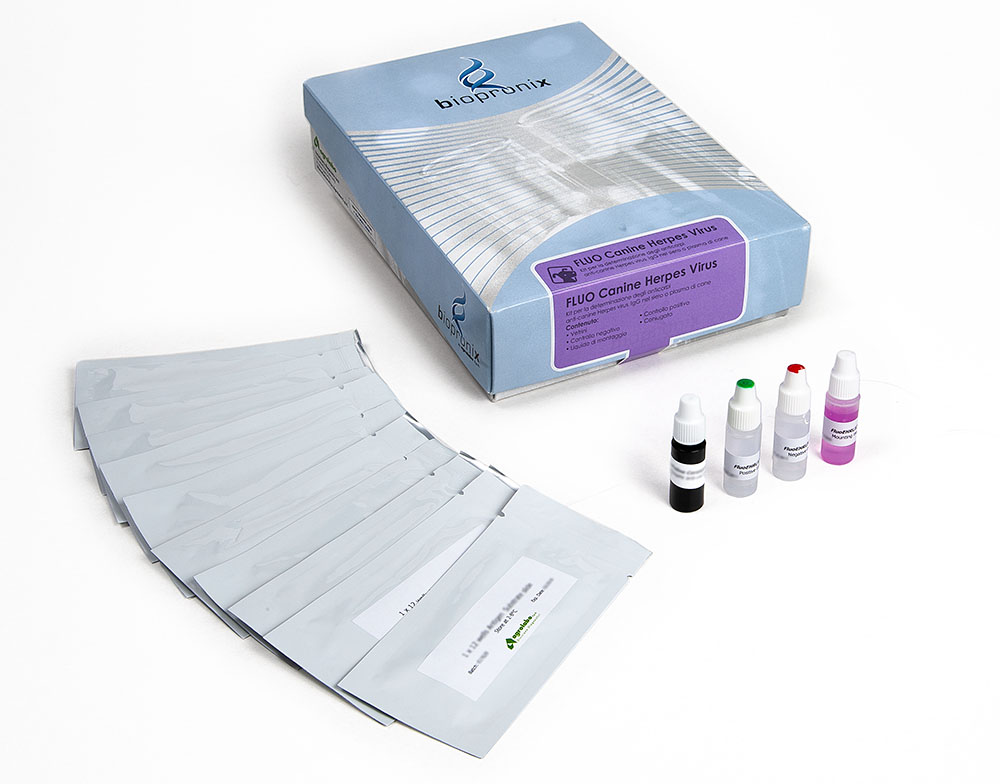
IFA kit for the detection of anti-Canine Herpesvirus IgG antibodies
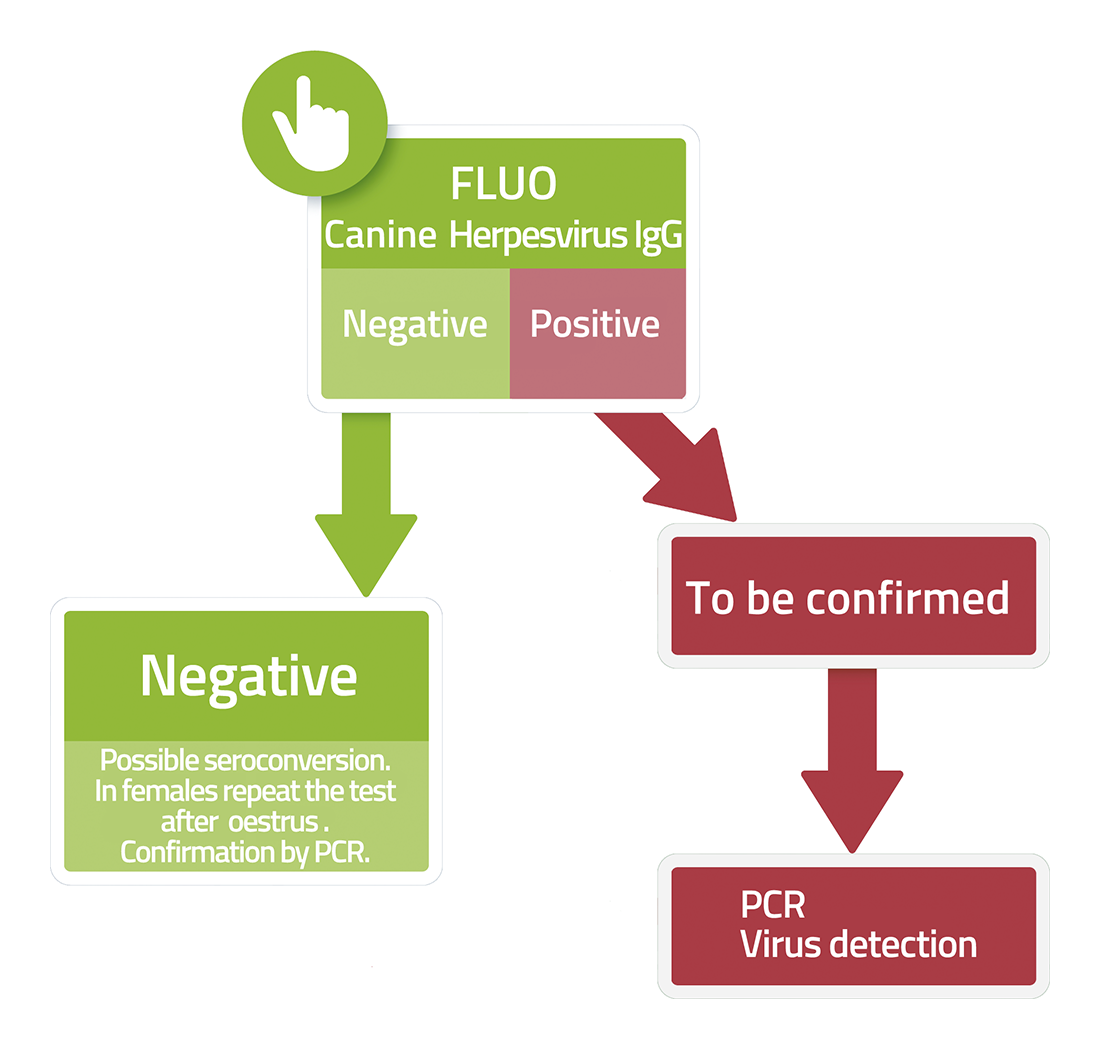
Fluo CANINE HERPESVIRUS is a test based on the immunofluorescence technique for the detection of IgG antibodies to Canine Herpesvirus (CHV) in dog serum or plasma samples.
Canine Herpesvirus 1 (CHV-1) is traditionally known in veterinary medicine as the causative agent of Fading Puppy Syndrome, a condition characterized by high mortality in infected newborns.
Like other members of the Alphaherpesvirinae subfamily, CHV-1 has evolved sophisticated host adaptation mechanisms to modulate viral replication, evade the immune response, and establish a lifelong persistent infection.
1.Thermal Adaptation: The Role of Temperature in Pathogenesis
One of the most distinctive characteristics of CHV-1 is its sensitivity to temperature, a factor that profoundly influences the virus's biological behavior and the distribution of lesions.
- Thermal Optimum: CHV-1 exhibits optimal replication capacity at relatively low temperatures, generally around 33–35°C. As the temperature rises to approximately 38-39°C (100.4-102.2°F) or during febrile episodes, viral replication is severely limited. This phenomenon is related to the reduced efficiency of some processes necessary for the virus's replication cycle, including regulation of viral gene expression and viral particle assembly.
- Why newborn puppies are vulnerable: In puppies in the first weeks of life, thermoregulatory systems are not yet fully mature. The ability to maintain a stable body temperature is reduced, and exposure to cold environmental conditions can cause hypothermia. Under this condition, tissue temperatures can reach levels favorable for CHV-1 replication, allowing systemic spread of the virus, involving numerous organs and potentially lethal consequences.
- Mucosal persistence in adults: In immunocompetent adult dogs, body temperature generally limits systemic spread of the virus. After primary infection, CHV-1 therefore tends to replicate primarily in the upper respiratory and genital mucosa. This localization contributes to the possibility of transmission through respiratory and genital secretions and may be associated with CHV-1's involvement in the complex of canine infectious respiratory diseases.
2.Neuronal latency: the mechanism of long-term persistence
Like all herpesviruses, CHV-1 has the ability to establish a latency phase, during which the viral genome remains present in the body without leading to significant production of new viral particles. This strategy allows the virus to persist in the host even after the primary infection has resolved.
- Transport to nerve ganglia: After initial replication in the mucosa, the virus can reach peripheral nerve endings and migrate along axons via the intracellular microtubule transport system. Retrograde transport allows the viral genome to reach the cell bodies of neurons, with its primary localization in the trigeminal ganglia, associated with respiratory/ocular infections, and in the sacral ganglia, associated with the genital tract.
- The viral episome and the molecular mechanisms of latency: Within the neuronal nucleus, CHV-1 DNA is not integrated into the host chromosome, but remains as a circular extrachromosomal element called an episome. During the latent phase, the virus drastically reduces the expression of genes required for replication and limits the production of viral proteins easily recognized by the immune system. During this phase, Latency-Associated Transcripts (LATs) and specific viral microRNAs, molecules involved in regulating gene expression and maintaining the latency state, are predominantly expressed.
3.Immune System Evasion: Inactivation of MHC-I
Physiological or pharmacological stressors, such as childbirth, hormonal changes, environmental stress, or corticosteroid treatment, can promote virus reactivation from the neuronal latent phase. In this phase, CHV-1 resumes active replication and migrates to peripheral tissues via anterograde axonal transport, reaching the mucosa, where it can be eliminated in secretions (viral shedding). To successfully complete this cycle, the virus must modulate several mechanisms of the cellular immune response.
- Modulation of antigen presentation: One of the body's main antiviral surveillance systems is represented by cytotoxic CD8+ T lymphocytes, which recognize and eliminate infected cells by recognizing viral protein fragments exposed on the cell surface by MHC class I molecules. Herpesviruses, including CHV-1, have evolved several mechanisms to interfere with this process.
- Interference with the TAP–MHC-I pathway: In infected cells, viral proteins are normally degraded into peptide fragments that are transported into the endoplasmic reticulum by the TAP (Transporter Associated with Antigen Processing) complex. There, the peptides are loaded onto MHC-I molecules, which expose them on the cell surface, allowing recognition by CD8+ T cells. CHV-1, like other alphaherpesviruses, can express proteins that interfere with this pathway, reducing the transport of viral peptides and limiting the correct expression of MHC-I on the cell surface. The result is a decrease in the visibility of the infected cell to the adaptive immune response, favoring the survival of cells hosting the virus during the replication phase.
- Balance between T cell evasion and NK cell control: Natural killer (NK) cells use the decrease in MHC-I as a signal to recognize altered or infected cells. Some alphaherpesviruses produce viral molecules capable of interacting with NK cell receptors or mimicking inhibitory signals associated with the presence of MHC-I, reducing cytotoxic activation.
4.The role of phagocytic cells in the spread of infection
In the most vulnerable individuals, such as newborn puppies or fetuses in cases of transplacental infection, CHV-1 can cause a systemic form characterized by the involvement of multiple organs. In addition to infection of epithelial cells, a role of cells of the phagocytic system, including tissue macrophages and antigen-presenting cells, has been hypothesized in the spread of the virus within the body.
- Intracellular survival and dissemination: Under physiological conditions, phagocytosis leads to the degradation of microorganisms through the fusion of the phagosome with lysosomes and the activation of intracellular antimicrobial mechanisms. Some viruses have developed strategies, promoting their own survival within host cells or altering cellular responses that would normally lead to their elimination. Involvement of immune cells can contribute to viral dissemination through the bloodstream to highly vascularized organs (liver, kidneys, lungs) and cause direct cellular damage, focal necrosis, and severe organ damage.
Bibliography
- Carmichael, L. E., et al. (1969) Temperature as a factor in resistance of young puppies to canine herpesvirus. Journal of Infectious Diseases, 120(6), 669-678.
- Gadsden, B.J., et al. (2012) Fatal Canid herpesvirus 1 infection in an adult dog. J Vet Diagn Invest. 24(3):604-7.
- Griffin, B.D., et al. (2010) Herpesviruses and immunity: the art of evasion. Vet Microbiol. 143(1):89-100.
- Ledbetter, E. C. (2013) Canine herpesvirus-1 ocular diseases of mature dogs. New Zealand Veterinary Journal, 61(4), 193-201.
- Miyoshi, M., et al. (1999) Detection of canine herpesvirus DNA in the ganglionic neurons and the lymph node lymphocytes of latently infected dogs. The Journal of Veterinary Medical Science, 61(4), 375-379.
- Ronsse, V., et al. (2005) Canine herpesvirus-1 (CHV-1): clinical, serological and virological patterns in breeding colonies. 64(1):61-74.
The severe form was found only in puppies under the age of one month, while the outcome is lethal only in subjects under the age of 2 weeks. In puppies, the disease begins between the 5th and 14th day of birth and presents with the following symptoms:
- emission of soft, odorless, yellow-green stools
- anorexia
- wheezing
- abdominal pain.
.
The course is rapid, death occurs in 24-48 hours.
In adult dogs the disease is limited to the reproductive organs. In females, a genital infection with multiple lymphoid nodules occurs. In males, preputial exudation of a serous character is observed between the 3rd and 7th day of exposure to the virus. The disease often starts in such a mild form that it can remain unnoticed.